Ronny Nienhold, et al, Nature Communications, 2020
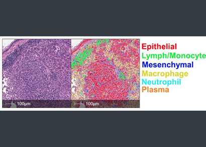
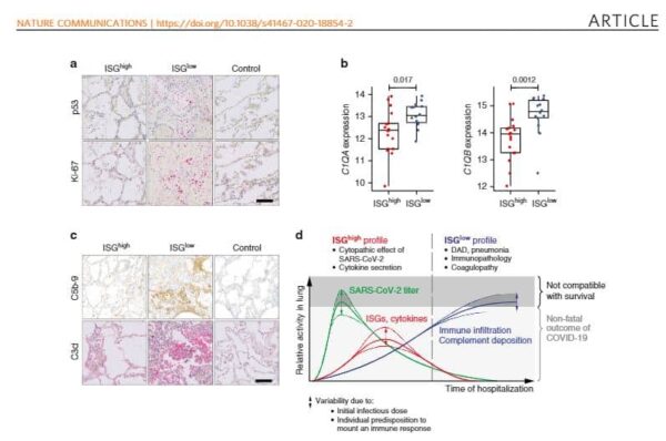
An international consortium of researchers characterized lung tissue from patients with COVID-19 using transcriptomic, histologic, and cellular analyses. Nienhold and colleagues report two phenotypes associated with lethal COVID-19 disease. One showed high levels of interferon stimulated genes in the lungs as well as limited lung damage and high levels of cytokines and viral loads. The second phenotype included severe lung damage with low levels of interferon stimulated genes, low viral loads, and high levels of CD8+ T cells and macrophages. As patients with the first phenotype die sooner, this highlights the need for biomarkers to classify COVID-19 patients and potentially guide treatment. HALO AI was trained using annotations from a pathologist to identify lung tissue and the resulting output was confirmed by pathology review. HALO was also used for quantification of immunohistochemistry analysis of CD3, CD4, CD8, CD20, CD68, CD123, CD163, and PD1.