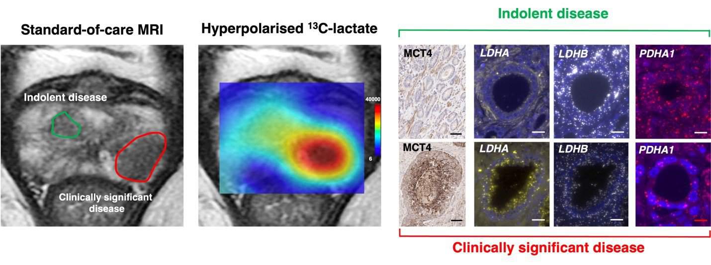
Dissecting Prostate Cancer Metabolic Compartmentalization Using Digital Pathology and Hyperpolarized MRI
Date: 6 April 2023
Time: 8:00 – 9:00 PST | 11:00 – 12:00 EST | 16:00 – 17:00 GMT
Location: Webinar
Learn how HALO® image analysis coupled with hyperpolarized MRI is advancing prostate cancer research.
Summary
Learning Objectives
- Learn how to use HALO Tissue Classifier module to spatially resolve different tissue subtypes in human prostate cancer
- Learn how to use HALO Membrane module to extract cell-type-specific membrane transporter expression patterns
- Learn how to use HALO FISH module to analyze cell-type-specific spatial transcriptomics data
Presenters

Nikita Sushentsev
PhD Graduate Student,
University of Cambridge, UK
Nikita is a PhD Student and Gates Scholar at the Department of Radiology, University of Cambridge. A medical doctor by training, Nikita is interested in biological validation and clinical translation of metabolic imaging techniques to address important diagnostic challenges in prostate cancer care. During his PhD, Nikita has demonstrated the potential of hyperpolarized 13C-MRI to non-invasively differentiate intermediate-risk prostate cancer based on their metabolic phenotype. To do so, he has deployed a variety of spatially resolved biological techniques including immunohistochemistry, spatial transcriptomics, and metabolomics. After his PhD, Nikita will continue his research in Cambridge as a Research Fellow of Emmanuel College.

Cara Brodie
Senior Scientific Associate Cancer Research UK Cambridge Institute
University of Cambridge, UK
Cara has a BSc Biological Science from University of Central Lancashire and a MSc in Biomedical science from Greenwich University.
Cara joined CRUK-Cambridge Institute (CI) Histopathology/ISH core facility in 2011 as a Scientific Officer developing her histopathology knowledge. Cara is now a Senior Scientific Associate for scanning and image analysis. The facility offers brightfield scanning with the Aperio AT2 (Leica) and fluorescent scanning of whole slides with both the Zeiss AxioScan Z1 and the PhenoImager HT (Akoya). Analysis of these images can be done in house or training is offered with the HALO software (Indica Labs).