


The Breast IHC Tumor Detection App is a pre-trained HALO AI classifier designed to detect, segment, and quantify tumor and other area across hematoxylin and DAB-stained whole-slide digital images of breast cancer.
Learn More
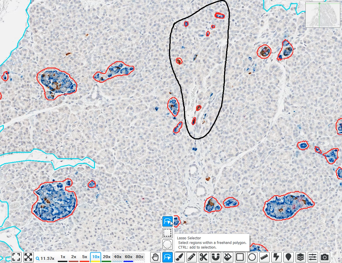
The NSCLC IHC Tumor Detection App is a pre-trained HALO AI classifier designed to detect, segment, and quantify tumor area and non-tumor area across hematoxylin and DAB-stained whole-slide digital images of NSCLC.
Learn More
The NSCLC IHC Cancer Cell Phenotyper App is a pre-trained HALO AI object phenotyper designed to detect, segment, and quantify non-cancer cells, IHC-positive cancer cells and IHC-negative cancer cells across hematoxylin and DAB-stained whole-slide digital images of NSCLC.
Learn More
The Breast IHC Cancer Cell Phenotyper App is a pre-trained HALO AI object phenotyper designed to detect, segment, and quantify cancer cells and other cells across hematoxylin and DAB-stained whole-slide digital images of breast cancer.
Learn More
The NSCLC H&E Cancer Cell Phenotyper App is a pre-trained object phenotyper designed to detect, quantify, and segment cancer cells from non-cancer cells across H&E-stained whole-slide digital images of NSCLC.
Learn More
The CRC H&E Cancer Cell Phenotyper App is a pre-trained HALO AI object phenotyper designed to detect, segment, and quantify cancer cells and other cells across H&E-stained whole-slide digital images of colorectal cancer.
Learn More
The Pan-Cancer H&E Lymphocyte Cell Phenotyper App is a pre-trained HALO AI object phenotyper designed to detect and quantify lymphocytes across whole slide H&E-stained images of multiple tumor types.
Learn More
The Gastric H&E Tumor Tissue Detection App is a pre-trained HALO AI masking classifier designed to segment tumor, stroma, necrosis/other, and glass areas across H&E-stained whole slide images of gastric cancer.
Learn More
The Head & Neck Squamous Cell Carcinoma (HNSCC) H&E Tumor Tissue Detection App is a pre-trained HALO AI masking classifier designed to segment tumor, stroma, necrosis/other, and glass area across H&E-stained whole slide HNSCC images.
Learn More
The Non-Small Cell Lung Cancer (NSCLC) H&E Tumor Tissue Detection App is a pre-trained HALO AI masking classifier designed to segment tumor, stroma, necrosis/other, and glass area across H&E-stained whole slide images of NSCLC.
Learn More
The Ovarian H&E Tumor Tissue Detection App is a pre-trained HALO AI masking classifier designed to segment tumor, stroma, necrosis/other, and glass area across H&E-stained whole slide images of ovarian cancer.
Learn More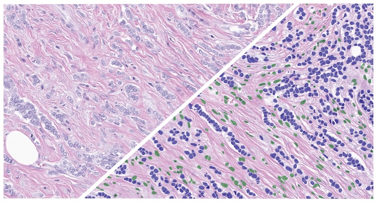
The Breast H&E Cancer Cell Phenotyper App is a pre-trained HALO AI phenotyper designed to detect and quantify cancer cells across whole slide H&E-stained images of breast cancer tissue.
Learn More
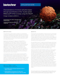
Characterizing the TME using IMC and HALO® Image Analysis
See in this app note how automated analysis of highly multiplexed IMC images using HALO and HALO AI yields rich cellular and spatial data from a streamlined workflow.
COMET™ and RNAscope™ Image Analysis Using HALO and HALO AI
Read this collaborative app note to learn how our Pharma Services team leveraged the HALO FISH-IF and Spatial Analysis modules and HALO AI for analysis of COMET™ and RNAscope™ images.
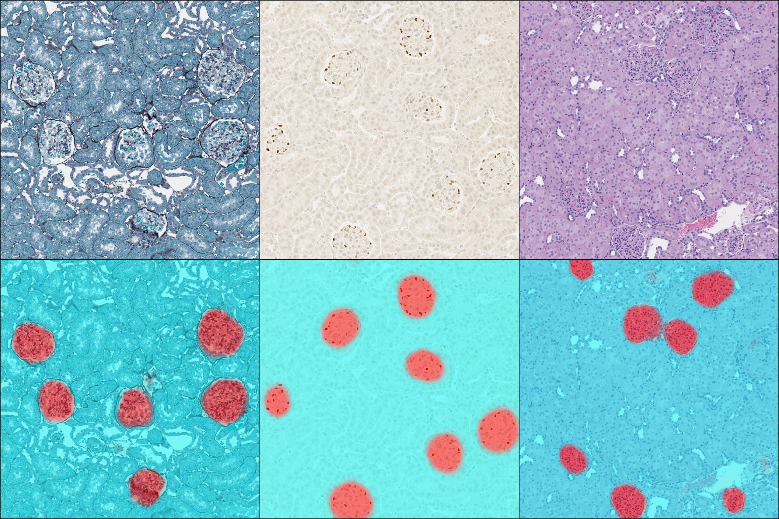
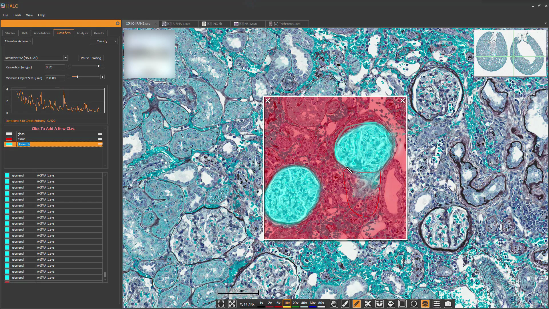
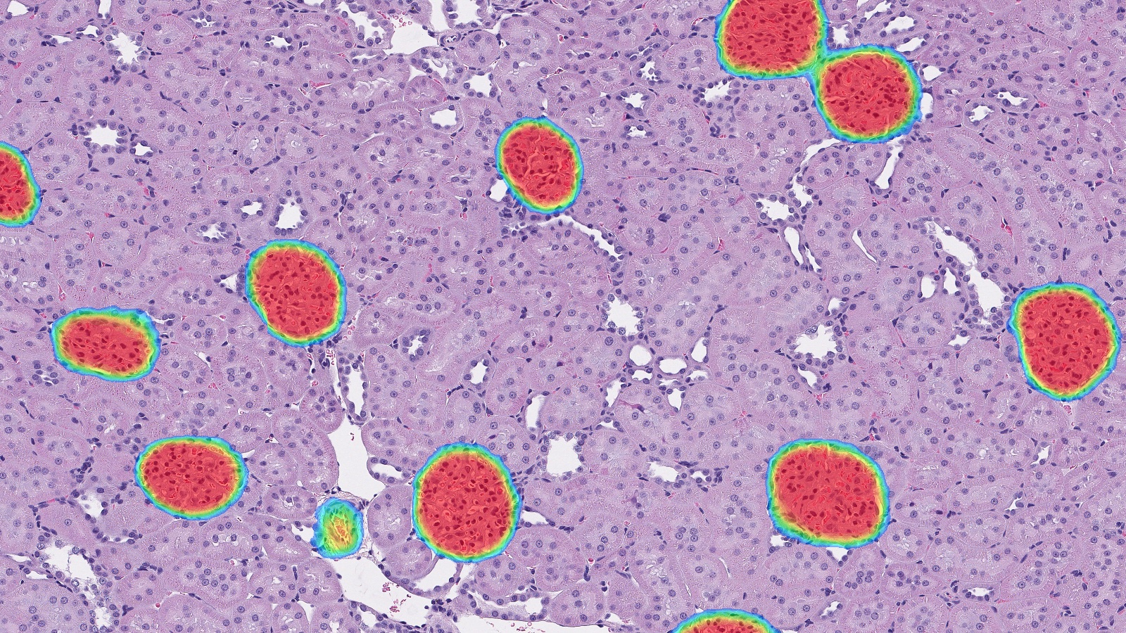
Glomeruli Quantification and Characterization Application Note
Don’t let varied stain selections hold back your digital image analysis of kidney sections! With HALO AI you can develop a tissue classifier that accurately detects and segments glomeruli in sections across a range of stains including H&E, trichrome, PAMS, and DAB with hematoxylin.
HiPlex RNAscope™ Application Note
Using our FISH module we demonstrate how to perform 12-plex RNAscope image analysis using ACD’s HiPlexv2 assay.

RNAscope Image Analysis Using HALO and HALO AI
Using our ISH module for brightfield and our FISH module for fluorescence, we demonstrate how to quantify ISH signal from RNAscope® assays on a per cell or per unit area basis and output an overall expression score based on ACD Bio’s recommended scoring guidelines.
HALO Platforms for Life Sciences Brochure
Download our brochure to learn about our HALO, HALO AI, and HALO Link platforms, and how they can advance your digital pathology image analysis and management workflows.
HALO, HALO AI, & HALO Link Cloud Services Brochure
Discover how our Cloud Services team can provide a secure, scalable, and flexible cloud deployment of HALO, HALO AI, and HALO Link.
Cloud Services and AI Diagnostics Case Study
Learn how the optimized, scalable infrastructure provided by Indica Labs Cloud Services enables the rapid development and robust collaboration of our AI Diagnostics group, powering the next generation of automated AI-based decision support tools.
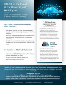
HALO in the Cloud at the University of Washington
Read this case study to learn how partnering with our Cloud Services team has delivered greater scalability and cost efficiency for the BioRepository and Integrated Neuropathology laboratory at University of Washington Medicine.
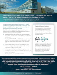
NCI Case Study
Download this case study to learn about considerations the National Cancer Institute made when planning to migrate its HALO, HALO AI, and HALO Link deployments to the cloud, and benefits the NCI experienced after migration, including 50% faster HALO image analysis.

Clustering Workflow eBook
Check out our collaborative eBook with Lunaphore to learn about an optimized workflow for extracting information from hyperplex datasets and illuminating changes in cellular neighborhoods using HALO, HALO AI, and the Lunaphore COMET platform.
Lunaphore COMET Hyperplex IF and HALO® Image Analysis eBook
Our collaborative eBook with Lunaphore shows how HALO and HALO AI, alongside the Lunaphore COMET instrument, enable a high-throughput workflow for phenotypic and spatial analysis of cells in the tumor microenvironment.
Qualitative and Quantitative Evaluation of the TME eBook
Check out this ebook to see how AI-based nuclear segmentation and tissue classification can streamline cell phenotyping across tissue compartments and samples.
Sequential Same Slide mIF and H&E eBook
Reveal more data on a single slide with this workflow for sequential same slide multiplex IF and H&E staining and imaging using HALO and HALO AI.
AI-Assisted Quality Control for Artifact Detection: Deployment in Image Management System with GLP Support
Download our poster to learn how artifact detection with the AI-powered SlideQC BF can advance your quality control pipelines with the GLP support of HALO Link.
All-In-One Digital Pathology: Compare and Contrast the Tumor Microenvironment of Renal Cell Carcinoma Tissue with Paired, Patient Derived Tumoroids
Check out this collaborative poster to learn about a streamlined triplex chromogenic workflow leveraging HALO and HALO AI for analysis of slides assayed with Leica Biosystems’ ChromoPlex III Triple Detection RUO.
An Automated Deep Learning Artifact Detection Tool for Quality Control of Whole-Slide Digital Pathology Images
Explore SlideQC: an AI-based tool which identifies pre-analytic processing errors in whole-slide images for quality control in digital pathology laboratories.
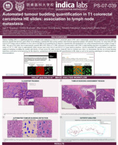
Automated Tumor Budding Quantification in Colorectal Carcinoma H&E Images
Learn about quantification of tumor budding in colorectal carcinoma using HALO and HALO AI.
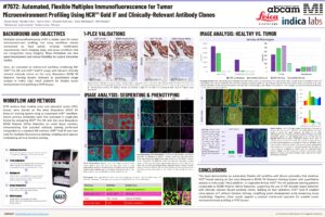
Automated, Flexible Multiplex Immunofluorescence for Tumor Microenvironment Profiling Using HCR Gold IF and Clinically-Relevant Antibody Clones
Check out this collaborative poster to learn about a streamlined triplex chromogenic workflow leveraging HALO and HALO AI for analysis of slides assayed with Leica Biosystems’ ChromoPlex III Triple Detection RUO.
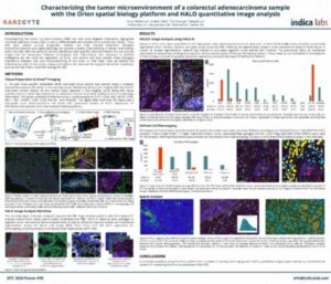
Characterizing the TME of a Colorectal Adenocarcinoma Sample with the Orion Spatial Biology Platform and HALO Image Analysis
Download our collaborative poster with RareCyte to learn how to analyze a colorectal adenocarcinoma sample with HALO and HALO AI
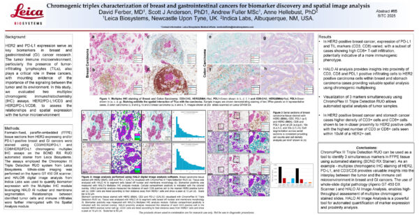
Chromogenic triplex characterization of breast and gastrointestinal cancers
Check out this collaborative poster to learn about a streamlined triplex chromogenic workflow leveraging HALO and HALO AI for analysis of slides assayed with Leica Biosystems’ ChromoPlex III Triple Detection RUO.
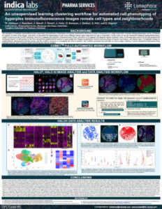
Clustering Workflow for Advanced Cell Phenotyping of Hyperplex IF Images
Download our collaborative poster with Lunaphore to learn about a clustering workflow that uses unsupervised learning for COMET™ images.
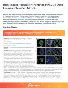
HALO AI High Impact Publications
Review this selection of high-impact publications for examples of HALO AI applications in fields ranging from metabolism to immuno-oncology and myology.
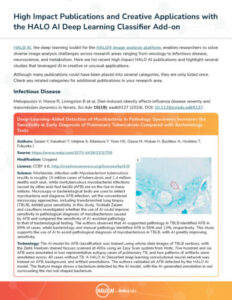
HALO AI Publications and Creative Applications
Check out this publication list to find recent high impact HALO AI publications and highlights of several studies leveraging AI in creative or unusual applications.
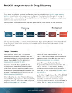
HALO Image Analysis in Drug Discovery
Review this publication list to learn how HALO platforms are advancing image analysis and helping accelerate findings throughout the drug discovery and development pipeline.


Fetal growth restriction (FGR) is a significant concern in obstetrics, affecting 5–10% of pregnancies globally. FGR is a major contributor to perinatal morbidity and mortality and poses a significant maternal risk due to potential co-occurrence with pre-eclampsia. Despite their impact,...
Learn More
This study examined CD8+ cell distribution in hepatocellular carcinoma (HCC) and peritumoral liver tissue to investigate the overall survival (OS) and recurrence-free survival (RFS). It is well understood that CD8+ lymphocytes are involved in both the anti-tumor response and in...
Learn More
EZH2 is the catalytic component of Polycomb Repressive Complex 2 (PRC2) and performs trimethylation of histone H3 at lysine 27 (H3K27me3) to silence chromatin. PRC2 is known to play different roles in different cancers and inhibitors of PRC2 histone methyltransferase...
Learn More
HALO AI Whitepaper
Check out this white paper for an in-depth introduction to the deep learning-based add-on to HALO, including available networks, common functions, and the “train – validate – test” Validation workflow.
Pharma Services and HALO Link White Paper
Download our white paper to learn how Indica Labs’ Pharma Services collaborates with customers and delivers HALO and HALO AI image analysis using HALO Link.
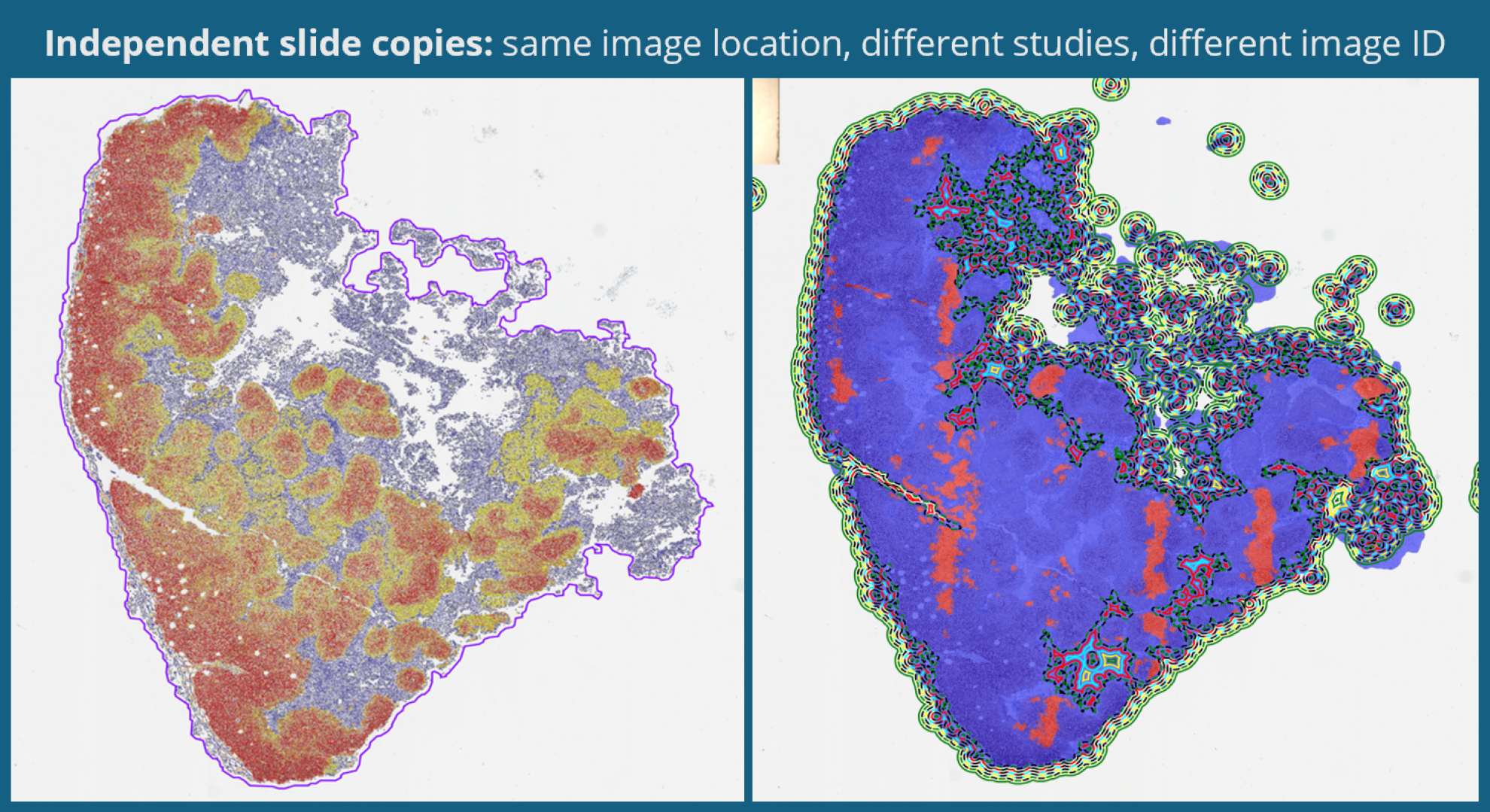
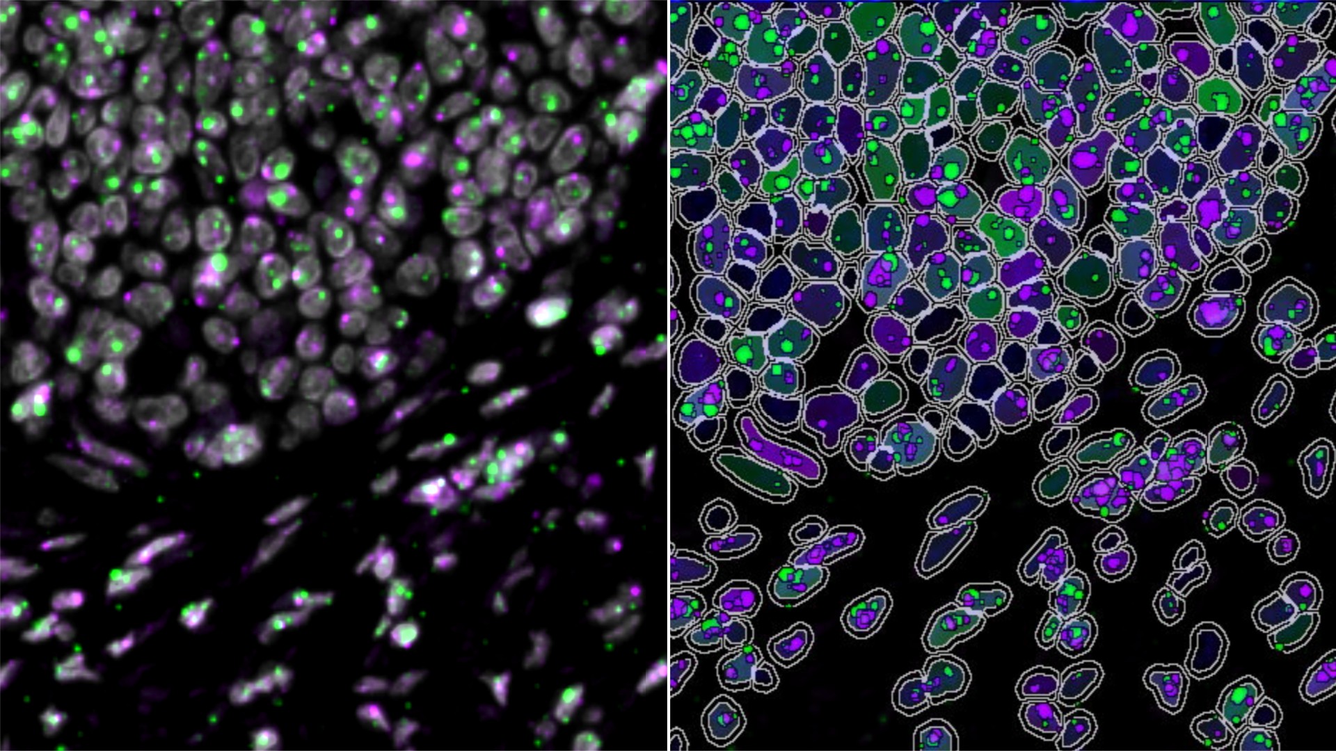
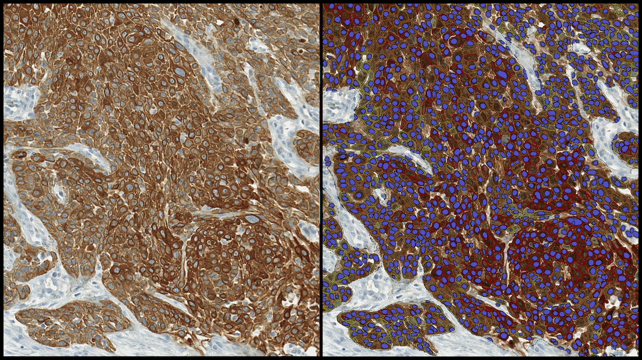
Quantitative Multiplex Chromogenic Image Analysis Guide
From stain and chromogen selection to honing nuclear and membrane detection in HALO, this comprehensive guide provides best practices and optimization techniques to help take your multiplex chromogenic image analysis to the next level.

Quantitative RNAscope™ Image Analysis Guide
From experimental design considerations to optimized setup of HALO image analysis parameters, our guide will help take your quantitative RNAscope™ image analysis to the next level.
What is new in HALO®, HALO AI, and HALO Link 4.3?
11 June 2026 | Learn about the exciting new features in the 4.3 versions of HALO, HALO AI, and HALO Link!
From Images to Insights: Getting Started with RNAscope™ Image Analysis in HALO®
25 June 2026 | Discover commonly used tools to get started today with RNAscope™ image analysis in HALO®
Getting Started with AI-Powered HALO Image Analysis
23 April 2026 | Learn how HALO and HALO AI enable quantitative image analysis, turning complex tissue data into actionable insights
Masterclass: Unlock Cell Population Discovery Using HALO® High Dimensional Analysis
31 March 2026 | Learn how unbiased high-dimensional analysis can be seamlessly combined with AI-powered HALO workflows for multiplex images!
Indica Labs Surpasses 3,000 Deployments of HALO Digital Pathology Software Worldwide Since 2013
Albuquerque, NM, March 3, 2026 – Indica Labs, the global leader in AI-powered digital pathology software and services, today announces surpassing the 3,000th installation of

Indica Labs’ Pharma Services Launches GCP-Compliant Image Analysis Service
Albuquerque, NM, July 8, 2025 – Indica Labs, the global leader in AI-powered digital pathology software and services, today announces that their Pharma Services team

Indica Labs Joins the AWS Partner Network
Albuquerque, NM, December 4, 2024 – Indica Labs, the global leader in AI-powered digital pathology software and services, announced today that they have joined the

The Institute of Molecular Pathology and Immunology of the University of Porto (IPATIMUP) Selects Indica Labs’ HALO AP® to Deliver AI-enabled Diagnostics
Albuquerque, NM, October 30, 2024 – The Institute of Molecular Pathology and Immunology of the University of Porto (IPATIMUP), among the leading institutes for clinical

Looking Back: Reviewing 2025 at Indica Labs
As we reach the end of 2025, the Indica Labs team extends our sincere thanks to the customers and collaborators who helped make this year
Advancing Neuroscience Research with HALO® and HALO AI
As scientists work to continue unraveling the structural, cellular, and molecular complexities of the brain and its diseases, the characteristics of this unique tissue pose
Unlocking the Potential of Antibody-Drug Conjugates (ADC) with HALO® Image Analysis and Indica Labs Pharma Services
Antibody-drug conjugates (ADC) represent one of the most promising classes of next-generation cancer therapeutics, combining antibody-based targeting with the potency of chemotherapy.
HALO®, HALO AI, and HALO Link 4.2 Features and Functionalities
In this blog post, you can learn about some of the new features in the 4.2 release, when to expect your chance to upgrade, and

Indica Labs’ Boston HALO® User Group Meeting 2026
12 May 2026 | Indica Labs is pleased to announce our Boston HALO® User Group Meeting at Le Méridien Boston Cambridge on 12 May from

Indica Labs’ Frankfurt HALO® User Group Meeting 2026
17 March 2026 | Indica Labs is pleased to announce our Frankfurt HALO® User Group Meeting at The Hilton Frankfurt Airport on 17 March from


Indica Labs’ London HALO® User Group Meeting 2025
9 December 2025 | Indica Labs is pleased to announce our London HALO® User Group Meeting at Hilton London Metropole on 9 December from 12:00