Maximizing use of HALO® and HALO AI™ for a comprehensive image analysis for HUMAN Brain FFPE Tissue samples in Alzheimer’s Disease
Date: 25 May 2022
Time: 8:00 – 9:00 PST | 11:00 – 12:00 EST | 16:00 – 17:00 GMT
Location: Webinar
Learn how HALO and HALO AI are advancing neuropathology research at the UW Medicine Biorepository and Integrated Research (BRaIN) laboratory
Summary
In this webinar, Jeanelle Ariza Torres, Research Scientist in the Biorepository and Integrated Neuropathology (BRaIN) at the University of Washington Department of Laboratory Medicine and Pathology, will present various alternatives of combining HALO AI networks in combination of HALO modules for the analysis of several morphological cellular stains and pathological protein expressions in FFPE human brain tissue samples in Alzheimer’s disease and related dementias (ADRD).
Jeanelle Ariza Torres, is the BRaIN laboratory lead for neuropathology quantitative analysis efforts in whole slide images (WSI) in human brain tissue samples and the design of protocols for IHC and IF stains in fresh frozen and FFPE human brain tissue for optimal image analysis.
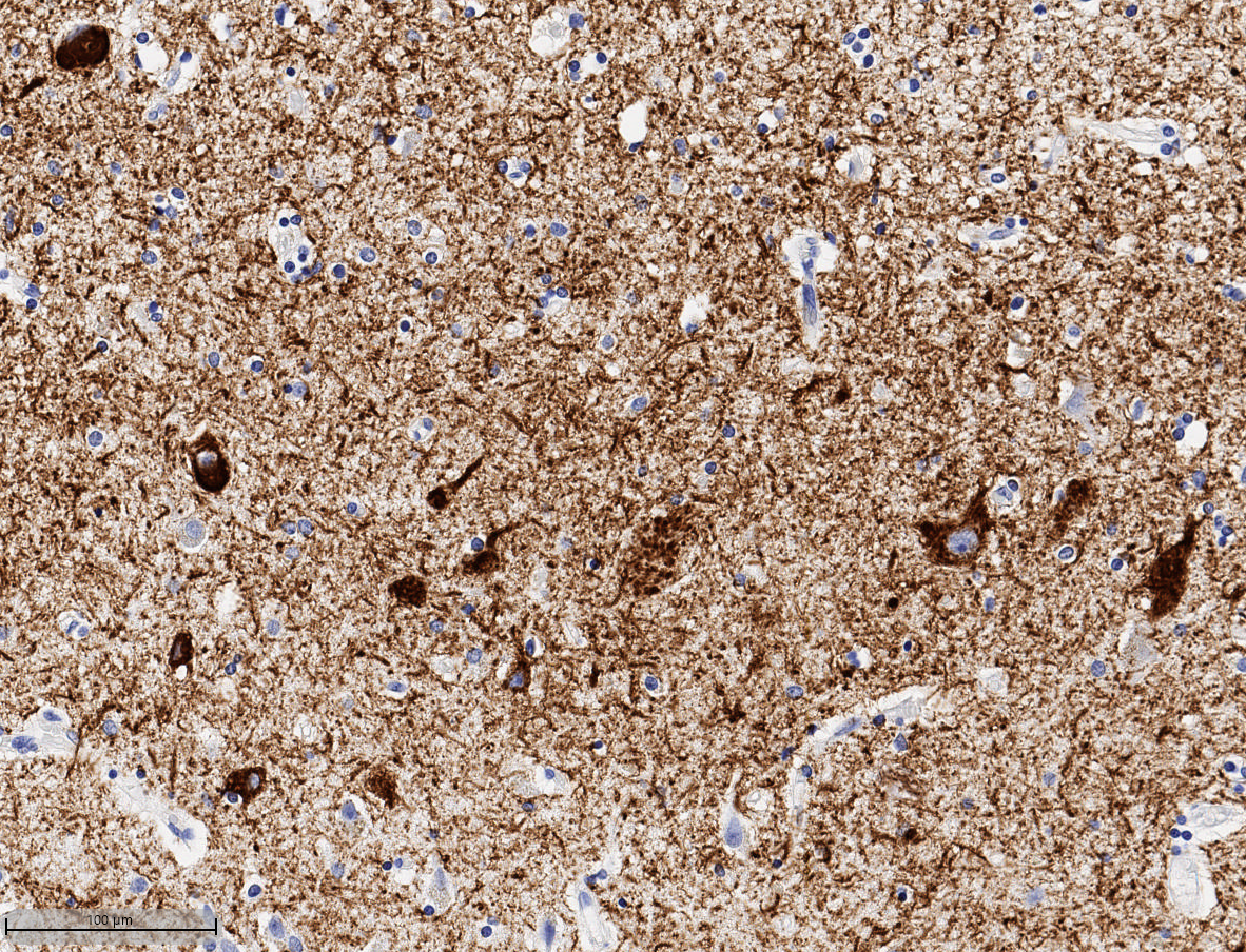
During this webinar, she will present different technical requirements focused on the single and duplex immunohistochemistry, as well as image analysis in Alzheimer’s disease FFPE human brain tissue samples. She will discuss various alternatives that can be used to train HALO AI for nuclear segmentation and ROI differential segmentation in the human brain cortex, and for morphometrical recognition for single cell markers in neurons (NeuN), astrocytes (GFAP) and microglia (IBA1 and pathological peptides including β amyloid (6e10) and cell bearing hyperphosphorylated TAU (AT8). Also, she will demonstrate different methods that can be used for image quality control and accurate analysis acquisition of minimal cell bearing immunoreaction in the form of intracellular inclusions for pathological peptides including phospho-TDP43 and α-synuclein to use it in combination with the Multiplex IHC module to obtain single cell information.
Finally, she will demonstrate strategies to get most out of the Multiplex IHC, Object Colocalization and Area Quantification modules with the goal of obtaining colocalization and co-expression information in double- IHC and single cell morphometric quantitative data.
Learning Objectives
- Provide important procedures to obtain the best quality sample in human brain tissue preparation for FFPE tissue sections.
- Demonstrate approaches to design single and double IHC for optimal image analysis in HALO.
- Learn how HALO AI networks can analyze brain tissue samples using cortical, and nuclear segmentation, QC in combination with HALO Area Quantification, Object Colocalization, Microglia and Multiplex IHC modules to obtain single object quantitative analysis information.
Presenters

Jeanelle Ariza Torres
Research Scientist, Neuropathology Keene Laboratory, Harborview Medical Center, University of Washington
University of Washington
Jeanelle Ariza Torres, a Research Scientist at the UW Medicine Biorepository and Integrated Research (BRaIN) laboratory led by C. Dirk Keene, MD, PhD, is working to perform and support research to understand normal brain anatomy and function and how this changes in injury and disease, such as Alzheimer’s disease (AD), Parkinson’s disease (PD), traumatic brain injury (TBI), chronic traumatic encephalopathy (CTE), and others
The BRaIN laboratory work supports the development of new treatments and preventive strategies to give those affected by brain injury and disease hope. The BRaIN laboratory, a leader in neuropathology research, is built to maximize the ability to study human donor research brain tissue. Dr. Keene and his team of researchers are actively studying and sharing thousands of samples each year with brilliant and innovative researchers at UW, in our region, and across the country – all working towards further understanding the effects of disease, injuries and possible cures.
Jeanelle leads the neuropathology quantification from image scanning, image storage and digital image effort using HALO software.
Jeanelle received her Biology degree, from the University Francisco Jose de Caldas in Bogotá Colombia and completed her post-bachelor studies in Forensic Anthropology from the University Nacional de Colombia.
Jeanelle has been working for 20 years in brain histology in neuroscience laboratories beginning at the Military University in Colombia and then in the US with the University of California San Francisco (UCSF) and University of California, Davis. For the past three years she has been working with the BRaIN laboratory at the University of Washington.
She has been working on the histology in neurodevelopment and neuropathology, understanding the differential cytoarchitectural brain organization from several animal species (mouse, rat, bat, snake, crocodile, catfish) and embryonic and adult human tissue brain samples
She has been in charge of the histological process, from tissue harvesting, storage, preparation, embedding in several mounting medias, histological stains, protocol design for immunofluorescence and immuno-enzymatic reactions for morphological cell identification and pathological protein identification for further image analysis using different software platforms.
Dr. C. Dirk Keene
Nancy and Buster Alvord Endowed Chair in Neuropathology
Professor, UW Department of Laboratory Medicine and Pathology;
Adjunct Professor, UW Departments of Ophthalmology and Neurological Surgery
Director, UW Neuropathology Division and Fellowship and Associate Director, UW Alzheimer’s Disease Research Center
Leader, UW BioRepository and Integrated Neuropathology (BRaIN) Laboratory and Precision Neuropathology Core
My overriding goal is to contribute knowledge and resources to normal human brain structure and function, brain aging, neurodegenerative disease, neurooncology, and neurotrauma research that lead to mechanistic discoveries and effective preventive and therapeutic strategies. I hope to accomplish this goal through neuropathological research and innovation, collaborative science, and education. I am Board Certified in Anatomic Pathology and Neuropathology, Direct the Division of Neuropathology for UW Medicine, have an active neurosurgical and ophthalmic neuropathology practice, and perform hundreds of comprehensive research brain autopsies for diverse clinical and research studies, including the University of Washington (UW) Alzheimer’s Disease Research Center (ADRC) and the Kaiser Adult Changes in Thought (ACT) study, the Seattle Longitudinal Study, the Pacific Northwest Brain Donor Network, and others. As Leader of the UW BioRepository and Integrated Neuropathology (BRaIN) Laboratory and Precision Neuropathology (NP) Core, I supervise a team whose primary goal is to respectfully and expeditiously perform brain autopsies in a manner that preserves tissues for diverse research applications under best practices conditions and protocols while providing accurate and timely neuropathologic diagnoses according to the latest guidelines. A principal goal for these efforts is to foster sensible sharing of these data and tissue resources with researchers to propel scientific discovery and best honor the generosity of the donor, his or her loved ones and caregivers, and their clinical research teams and studies. The primary goals for my research lab are to develop, deploy, and promote technologies that accentuate the scientific utility of archived and prospectively acquired human brain tissue, to utilize discoveries derived from these critical resources to develop and test hypotheses in experimental systems, and to apply this knowledge to develop therapeutic strategies.