Multi-omics analysis demonstrates repetitive head impacts induce neuronal loss and neuroinflammation in young athletes
Date: 21 October 2025
Time: 8:00 – 9:00 PDT | 11:00 – 12:00 EDT | 16:00 – 17:00 BST
Location: Webinar
Learn how HALO image analysis is helping to advance the understanding of chronic traumatic encephalopathy!
Summary
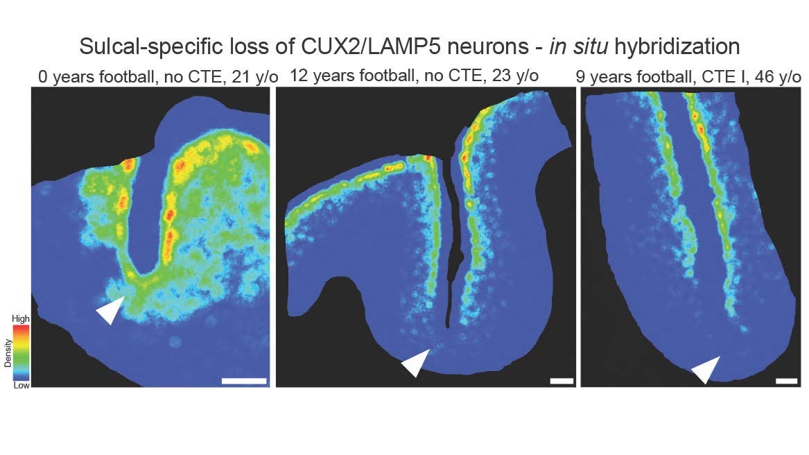
Repetitive head impacts (RHI) sustained from contact sports are the largest risk factor for chronic traumatic encephalopathy (CTE). Currently, CTE can only be diagnosed after death and the multicellular cascade of events that trigger initial hyperphosphorylated tau (p-tau) deposition remain unclear. Further, the symptoms endorsed by young individuals with early disease are not fully explained by the extent of p-tau deposition, severely hampering development of therapeutic interventions. Using genomics, spatial transcriptomics, and histology it was observed that RHI exposure associates with a multicellular response in young individuals (<51 years old) prior to the onset of CTE p-tau pathology that correlates with number of years of RHI exposure. SPP1+ inflammatory microglia, angiogenic and inflamed endothelial cell profiles, reactive astrocytes, and altered synaptic gene expression in excitatory and inhibitory neurons were identified in all individuals with exposure to RHI. Surprisingly, elevation in collagen deposition around blood vessels that might be related to long term disease outcomes was also observed. Overall, these results provide robust evidence that multiple years of RHI exposure is sufficient to induce lasting cellular alterations that may underlie p-tau deposition and help explain the early pathogenesis in young former contact sport athletes.
Learning Objectives
- Understand how HALO image analysis can assist in characterizing pathology in neurodegenerative diseases
- Understand how HALO AI can be used to identify unique cell populations with complex, varied shapes
- Explore how genomic data can be validated using histologic images analysis
Presenter

Jonathan Cherry, PhD
Research Health Scientist
VA Boston Healthcare System
Director, Digital Pathology Core & Research Neuropathologist
Boston University Alzheimer’s Disease Research Center and Boston University Chronic Traumatic Encephalopathy Center
Assistant Professor of Pathology and Laboratory Medicine
Boston University Avedisian & Chobanian School of Medicine
Dr. Cherry is an Assistant Professor of Pathology and Laboratory Medicine. He completed his undergraduate studies with a BS in biology at Ursinus College in 2008, earned his doctoral degree in Pathology from the University of Rochester in 2015, and joined the McKee laboratory as a postdoctoral researcher the same year. He was appointed as an assistant professor at Boston University School of Medicine in 2019. Dr. Cherry also holds a research health scientist position within the VA Boston Healthcare System. His laboratory space is located at the Jamaica Plain VA hospital where he performs his research and helps support the UNITE brain bank.
Dr. Cherry’s research interests focus on understanding how neuroinflammation after repetitive traumatic brain injury contributes to CTE pathogenesis. Specifically, Dr. Cherry seeks to identify what role microglia and inflammatory cytokines play in the early onset of hyperphosphorylated tau accumulation and spread.
Dr. Cherry’s research also extends to histology analysis and machine learning. He is the director of the BU digital pathology core. This research entails exploring novel ways to analyze human postmortem tissue using machine learning algorithms for neuropathologic targets and better characterize pathology across a spectrum of neurodegenerative diseases including Alzheimer’s disease, chronic traumatic encephalopathy, frontotemporal lobar degeneration, motor neuron diseases, and others.