Clearly Visualize Digital Pathology Images with View Settings Adjustments and the Figure Maker Tool
Whether a user’s goal is to accurately display subtle phenotypes in a multiplex fluorescence image or to build intuitive figures for a manuscript, accurately visualizing digital pathology images is critical. Additionally, the proliferation of highly multiplex imaging along with the increasing use of cloud-based collaborative platforms are placing new performance and accessibility demands on tools for optimizing and sharing images.
Designed to overcome these challenges and power your next discovery, our HALO® image analysis platform and browser-based HALO Link collaborative image management system include powerful capabilities for visualizing digital pathology images. In this blog we’ll explore these platforms’ tools for view settings adjustment for fluorescent images and the integrated Figure Maker tool. Together, these features equip researchers to accurately observe and convey the complexity of their samples and the details of their analysis.
Optimizing Visualization with View Settings Adjustment
For fluorescent image datasets, the ability to fine-tune image display parameters is essential for interpreting and presenting results, and both HALO and HALO Link include robust capabilities for adjusting view settings. These settings allow researchers to tailor image display in real time, optimizing visibility across signal intensities and tissue regions. Critically, adjustments do not alter the original image data or impact analysis outputs.
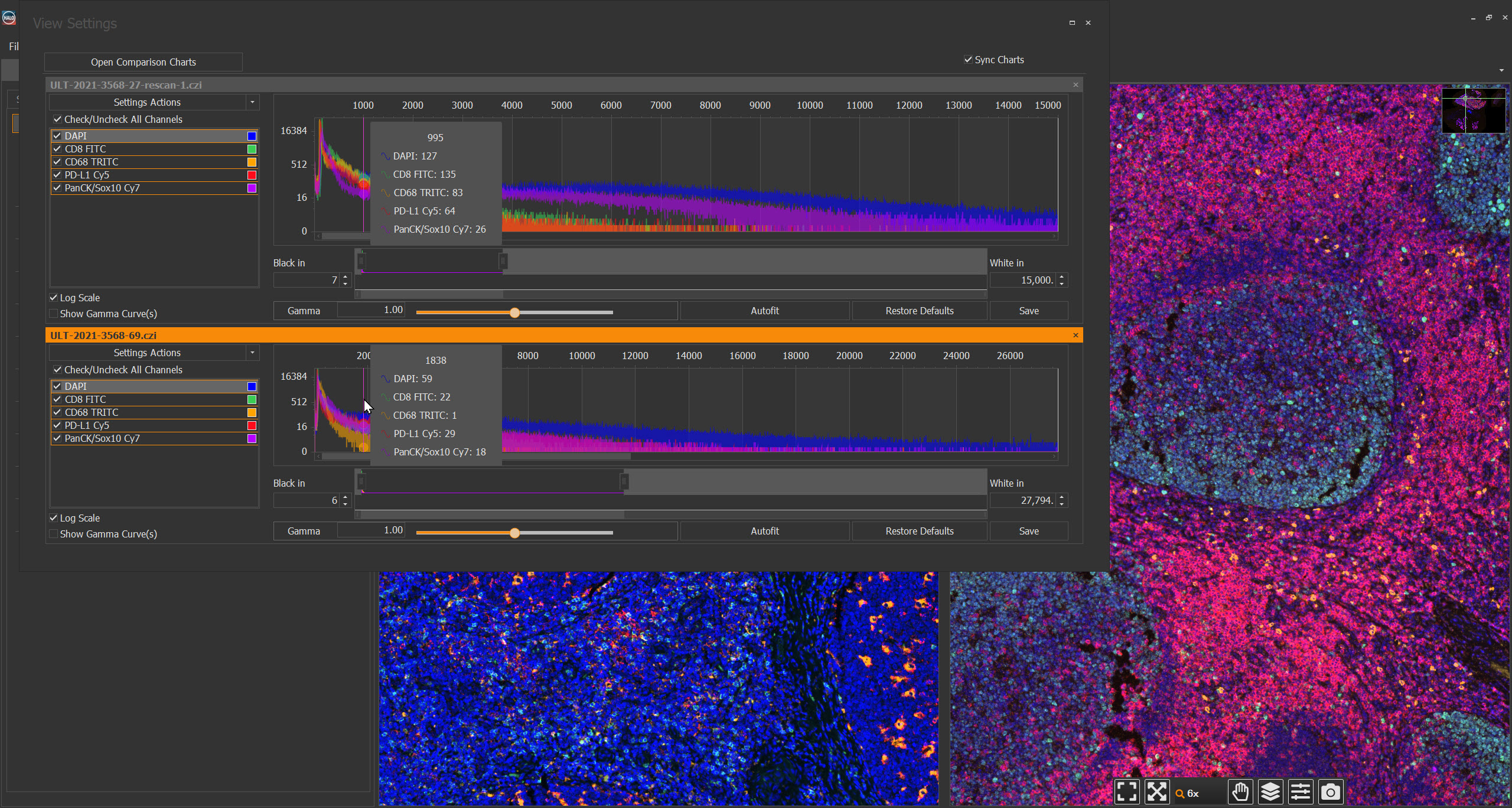
In both platforms, individual channels can be toggled on and off, renamed, assigned custom colors, and have their gamma value adjusted from the Channels tab. From this tab, users can open the View Settings window for a closer look at channel data and to further refine how channels are displayed. The View Settings window displays channel histograms and allows users to adjust black in and white in values for each channel.
HALO builds on these shared capabilities with the option to display histograms for up to three images side-by-side and synchronize histograms, facilitating comparison of channel intensity across images. Users can also leverage an autofit algorithm to set per-channel black in and white in values with a click, a time-saving feature that streamlines processing of large datasets.
To further assist with selecting view settings, HALO enables users to toggle on and off gamma curves for each channel as well as logarithmic scaling. After users have optimized their view settings, they can apply them in batch or export them to share with collaborators.
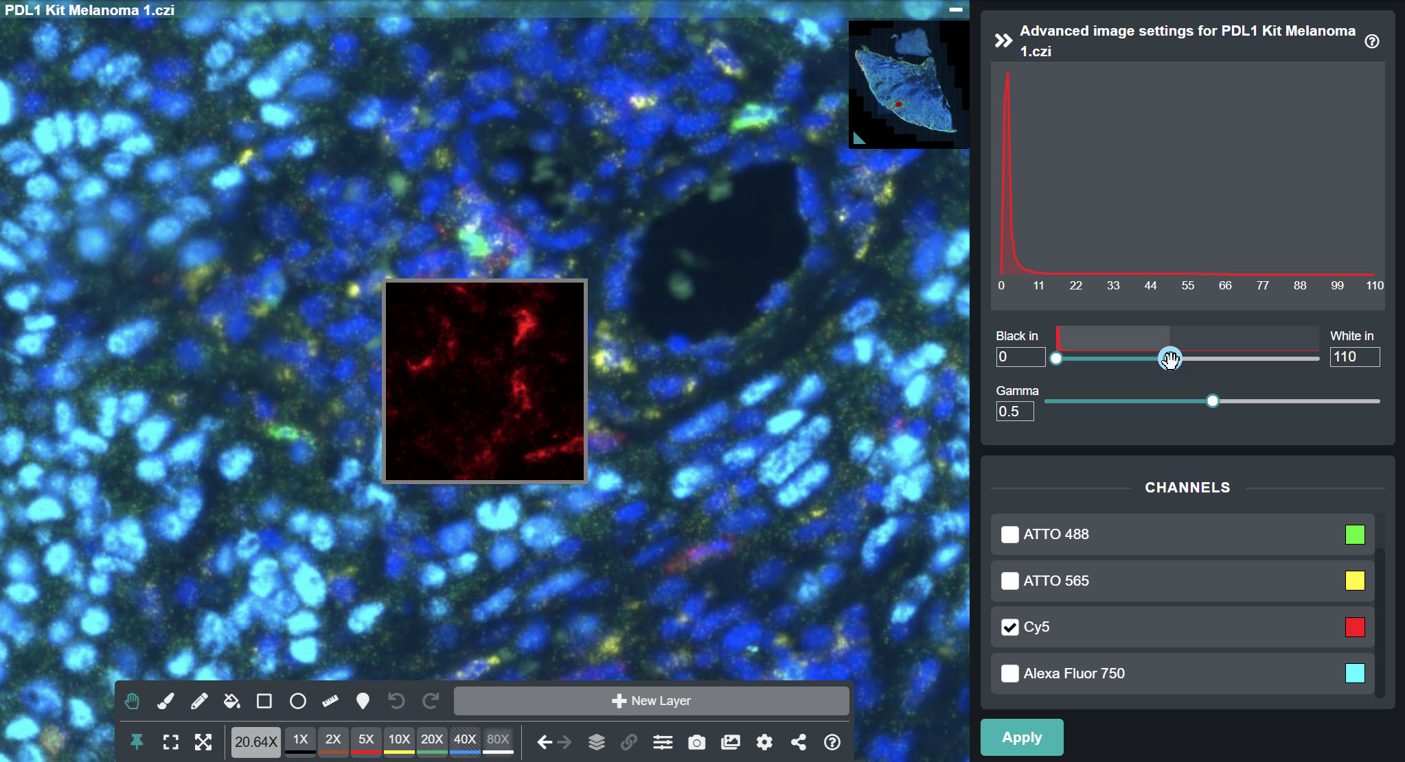
Beyond the core View Settings window capabilities, HALO Link offers a unique feature to enhance user experience in remote, browser-based environments: a live preview window. This tool enables real-time review of view settings changes before they are applied to the full image, limiting demands on compute and bandwidth resources.
Streamlined and Integrated Figure Creation
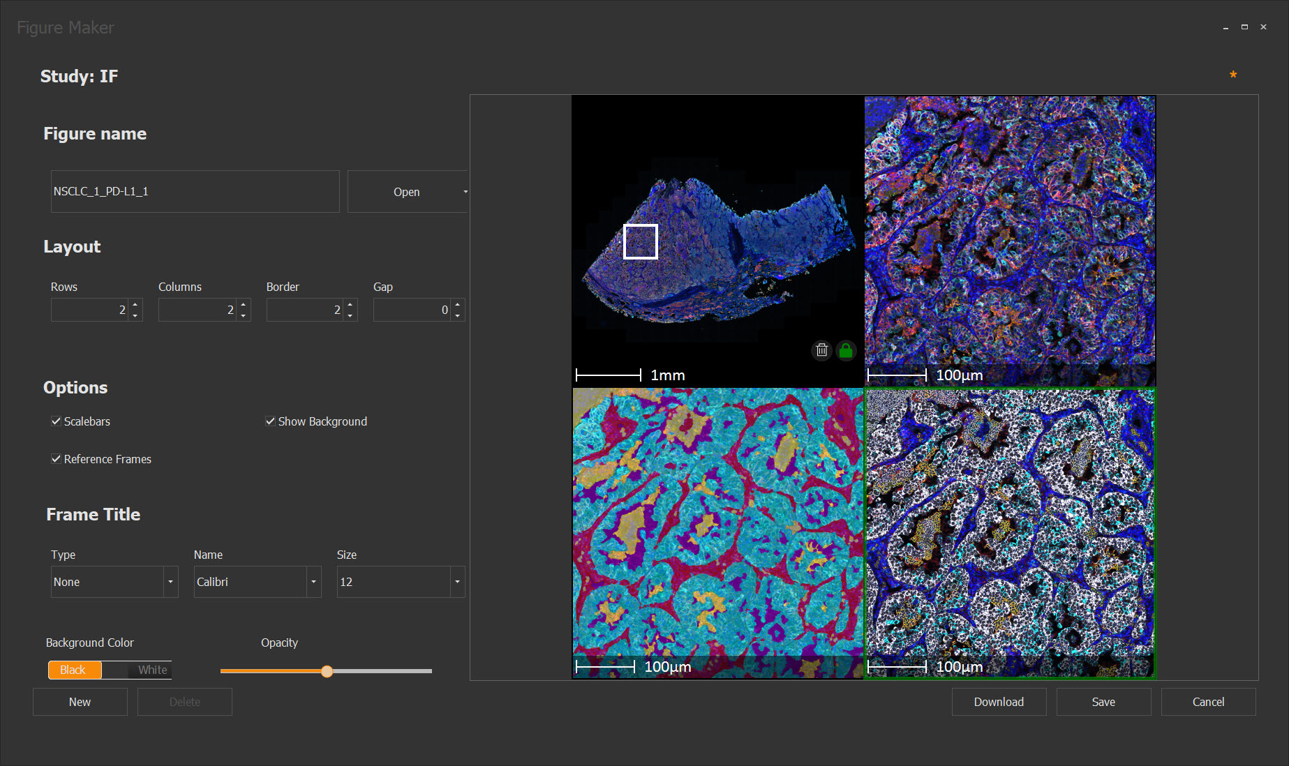
While adjusting image view settings is key to accurately visualizing digital pathology images within HALO and HALO Link, the Figure Maker tool enables researchers to go a step further by enabling the creation of high-quality, publication-ready figures directly within these platforms. Designed to streamline the figure preparation process, Figure Maker offers an intuitive interface for composing multi-panel layouts that clearly communicate key observations.
In both platforms, users can construct figures of up to 6×6 grids. The tool populates the active panel directly from the viewer’s current field of view, and changes, such as zooming, panning, toggling markups, or displaying annotations, are dynamically reflected.
After selecting their images, users can choose to include panel-level customizations including adjustable spacing and border thickness around each panel, panel labels, scalebars, and reference frames. Color and opacity adjustments for text and frame elements offer further flexibility. Once finalized, users can save figures to a study for future editing or download figures in multiple formats with customizable resolution and image scaling options.
Conclusions
From initial image review to publication of graphics that clearly, accurately, and efficiently communicate findings, ensuring optimal image display is foundational to digital pathology. With advanced view settings adjustments for fluorescent images and the integrated Figure Maker, HALO and HALO Link give researchers the tools they need to clearly observe and communicate their findings using streamlined, time-saving workflows.
If you’re interested in learning more about these and the other capabilities offered by HALO and HALO Link and scheduling a hands-on demo, contact us at info@indicalab.com.