


HALO® Analysis Applications in Neuroscience Research
15 August 2018 | In this one hour webinar, Indica Labs’ Application Scientist, Alyssa Myers, will discuss how the HALO platform can be used for

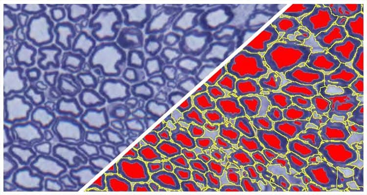
Simultaneously analyze up to five chromogenic stains and measure object density, area, diameter, and optical density, as well as colocalizations, if applicable.
Learn More
Simultaneously analyze an unlimited number of fluorescent dyes and measure object density, area, diameter, and intensity, as well as colocalizations, if applicable.
Learn More
Quantify microglial activation based on length and thickness of microglial processes.
Learn More