Measure microglia cell activation in brightfield images with the HALO® Microglial Activation module. This module detects microglia, soma, and processes, and reports the number of activated and inactivated microglial cells and the number of non-microglia cells detected. The tool also precisely measures cell processes, reporting the total process area and length as well as the average process area, length, and thickness, and quantifies process branching by counting the number of branch points and end points per cell. Leverage optional interactive markup images to dynamically explore your results by toggling markup of activated and inactivated microglia, microglial processes and skeleton, branch and end points, and microglia radius.
Try it out! Click here to initiate your free proof-of-concept HALO image analysis.
File formats supported by the HALO image analysis platform:
- Non-proprietary (JPG, TIF, OME.TIFF)
- Nikon (ND2)
- 3D Histech (MRXS)
- Akoya (QPTIFF, component TIFF)
- Olympus / Evident (VSI)
- Hamamatsu (NDPI, NDPIS)
- Aperio (SVS, AFI)
- Zeiss (CZI)
- Leica (SCN, LIF)
- Ventana (BIF)
- Philips (iSyntax, i2Syntax)
- KFBIO (KFB, KFBF)
- DICOM (DCM*)
*whole-slide images
Announcing the launch of a new Microglial Activation module for fluorescence and an updated brightfield module
In this blog post, you can learn about some of the new features in these modules, where to find the user guides, tutorial videos, and
Masterclass Webinar: Neurobiology Image Analysis with HALO and HALO AI
29 September 2022 | Join us for this 1-hour webinar for a live demonstration of neurobiology image analysis using HALO® and HALO AI. Dr. Levi
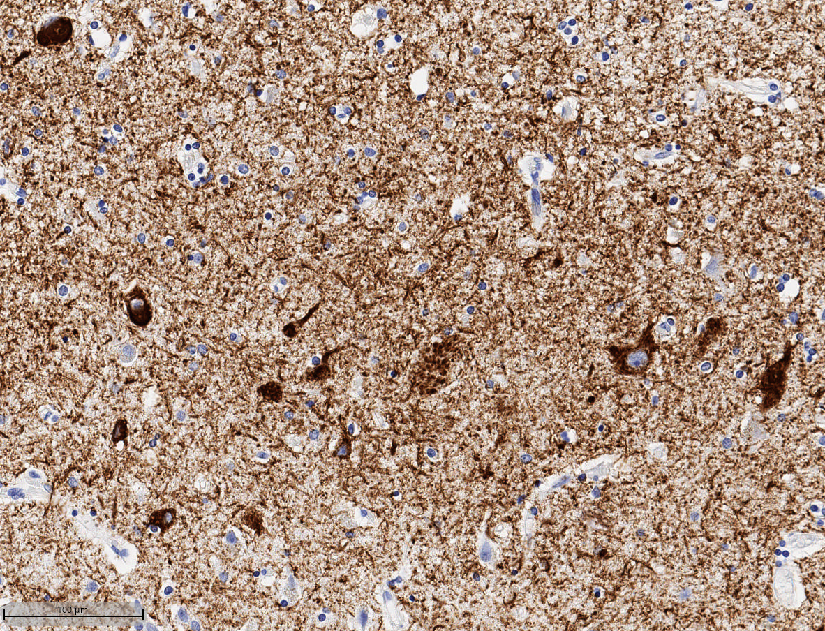
Maximizing use of HALO® and HALO AI for a comprehensive image analysis for HUMAN Brain FFPE Tissue samples in Alzheimer’s Disease
25 May 2022 | In this 60-min webinar, Learn how HALO and HALO AI are advancing neuropathology research at the UW Medicine Biorepository and Integrated
HALO® Analysis Applications in Neuroscience Research
15 August 2018 | In this one hour webinar, Indica Labs’ Application Scientist, Alyssa Myers, will discuss how the HALO platform can be used for
Publication Spotlight
The table below includes publications that cite the Microglial Activation modules.
Your publication not on the list? Drop us an email to let us know about it!
Title | Authors | Year | Journal | Application | HALO Modules | Product |
---|---|---|---|---|---|---|
Age-related pathology after adenoviral overexpression of the leucine-rich repeat kinase 2 in the mouse striatum | Kritzingerab A, Ferger B, Gillardon F, Stierstorfer B, Birk G, Kochanek S, Ciossek T | 2018 | Neurobiology of Aging | Neuroscience | Area Quantification, Object Colocalization, Microglia | HALO |
Diverse human astrocyte and microglial transcriptional responses to Alzheimerís pathology | Smith A, Davey K, Tsartsalis S, Khozoie C, Fancy N, Tang S, Liaptsi E, Weinert M, McGarry A, Muirhead R, Gentleman S, Owen D, Matthews P | 2021 | Acta Neuropathologica | Neuroscience | Area Quantification, Multiplex IHC, Microglia | HALO |
T Cells Limit Accumulation of Aggregate Pathology Following Intrastriatal Injection of†?-Synuclein Fibrils | George S, Tyson T, Rey N, Sheridan R, Peelaerts W, Becker K, Schulz E, Meyerdirk L, Burmeister A, von Linstow C, Steiner J, Galvis M, Ma J, Pospisilik J, Labrie V, Brundin L, Brundin P | 2021 | Journal of Parkinson's Disease | Immunology, Neuroscience | Microglia | HALO |
Traumatic Brain Injury Leads to Alterations in Contusional Cortical miRNAs Involved in Dementia | Naseer S, Abelleira-Hervas L, Savani D, de Burgh R, Aleksynas R, Donat C, Syed N, Sastre M | 2022 | Biomolecules | Neuroscience | Microglia | HALO |
Association between APOE genotype and microglial cell morphology | Kloske C, Gearon M, Weekman E, Rogers C, Patel E, Bachstetter A, Nelson P, Wilcock D | 2023 | Journal of Neuropathology & Experimental Neurology | neuroscience | Object Colocalization, Microglia | HALO |
Characterization of Traumatic Brain Injury in a Gyrencephalic Ferret Model Using the Novel Closed Head Injury Model of Engineered Rotational Acceleration (CHIMERA) | Krieg J, Leonard A, Tuner R, Corrigan F | 2023 | Neurotrauma Reports | Neuroscience | Microglia | HALO |
Anatomical distribution of Fyn kinase in the human brain in Parkinson's disease | Guglietti B, Mustafa S, Corrigan F, Collins-Praino LE | 2023 | Parkinsonism & Related Disorders | Neuroscience | Microglia | HALO |
Female mice display sex-specific differences in cerebrovascular function and subarachnoid haemorrhage-induced injury | Dinh DD, Wan H, Lidington D, Bolz SS | 2024 | EBioMedicine | Neuroscience | Microglia | HALO |
Related HALO Modules
Simultaneously analyze up to five chromogenic stains and measure object density, area, diameter, and optical density, as well as colocalizations, if applicable.
Learn MoreSimultaneously analyze an unlimited number of fluorescent dyes and measure object density, area, diameter, and intensity, as well as colocalizations, if applicable.
Learn MoreQuantify microglial activation in fluorescence based on detection of microglia, soma, and processes, by counting branch points, and by determining area, length, and thickness of processes.
Learn MoreWant to Learn More?
Fill out the form below to request information about any of our software products.
You can also drop us an email at info@indicalab.com