


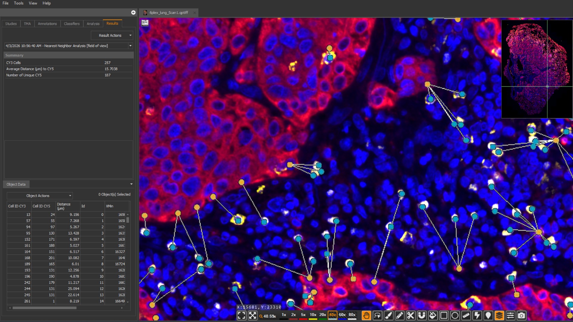
The Spatial Analysis module is a suite of four algorithms which identify proximity and relative spatial distribution of cells and objects across whole slide images, tissue boundaries, or serial sections. The Spatial Analysis module includes algorithms for nearest neighbor analysis, proximity analysis, infiltration analysis, and density heatmaps and can be used in conjunction with any cell-based analysis module for brightfield or fluorescence.
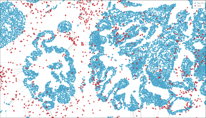
Nearest Neighbor
Determine the average distance and number of unique neighbors between any two cell or object populations using the nearest neighbor algorithm. Outputs include an image markup, a HALO Spatial Plot, and Object Pair Data.

Proximity Analysis
Determine the number of cells or objects within a certain distance of another object or cell with the proximity analysis algorithm. Outputs include an image markup, a proximity histogram, a HALO Spatial Plot, and Object Pair Data.
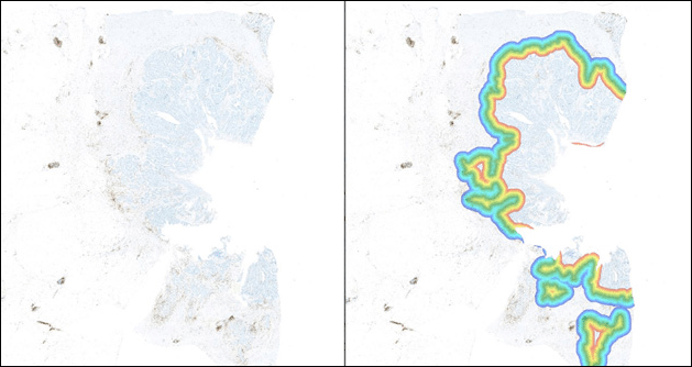
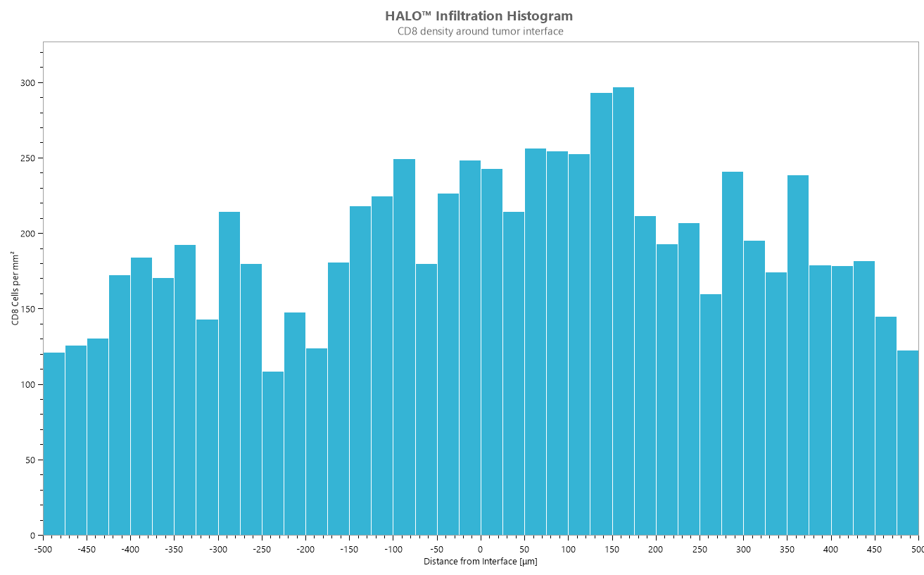
Infiltration Analysis
Determine the number of cells or objects within a set range of an annotated region of interest using the infiltration analysis algorithm. Outputs include an image markup, an infiltration histogram, a HALO Spatial Plot, and Object Pair Data.
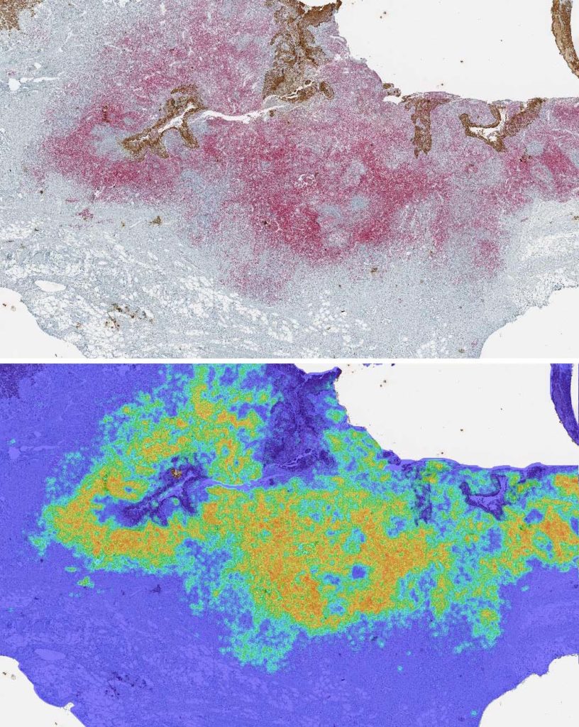
Density Heatmaps
The density heatmap algorithm measures the density of a selected cell or object population within a certain radius. Outputs include a markup image, Summary Data, and optionally, Object Data. Users also have the option to subdivide or bin Summary Data and can automatically generate output annotations.
Reach out to us at info@indicalab.com to learn more about the Spatial Analysis module.
- Non-proprietary (JPG, TIF, OME.TIFF) (JPG, TIF, OME.TIFF)
- Nikon (ND2)
- 3D Histech (MRXS)
- Akoya Biosciences/Quanterix (QPTIFF, component TIFF)
- Olympus / Evident (VSI)
- Hamamatsu (NDPI, NDPIS)
- Aperio/Leica Biosystems (SVS, AFI)
- Zeiss (CZI)
- Leica (SCN, LIF)
- Ventana/Roche (BIF)
- Philips (iSyntax, i2Syntax)
- KFBIO (KFB, KFBF)
- DICOM (DCM*)
*whole-slide images
Performing Spatial Analysis Workflows with HALO®
19 May 2026 | Learn how to unlock deeper insights and enhance your data using the HALO Spatial Analysis module

Masterclass Webinar: Performing Highly Multiplexed IF and Spatial Analysis Workflows with HALO®
25 June 2024 | Join us for this 1-hour webinar to see a live demonstration of the HALO® image analysis platform focused on highly multiplexed

A role for HALO® in characterizing cell heterogeneity across organ systems: from the liver to the brain
12 October 2023 | Please join us for this 1-hour webinar to learn about characterizing cell heterogeneity with HALO to gain new biological insights.
Advanced Multiplex and Spatial Analysis Methods using HALO Data
9 June 2022 | In this 90-minute webinar, Learn about an open-source MIBI image analysis pipeline using HALO data and how to validate a multiplex
Publication Spotlight
The table below includes publications that cite the Spatial Analysis module.
Your publication not on the list? Drop us an email to let us know about it!
| Title | Authors | Year | Journal | Topics | HALO Modules | Products |
|---|---|---|---|---|---|---|
| Neoadjuvant radioimmunotherapy in pancreatic cancer enhances effector T cell infiltration and shortens their distances to tumor cells | WANG J, GAI J, ZHANG T, NIU N, QI H, THOMAS II D L, LI K, XIA T, RODRIGUEZ C, PARKINSON R, DURHAM J, MCPHAUL T, NARANG A K, ANDERS R A, OSIPOV A, WANG H, HE J, LAHERU D A, HERMAN J M, LEE V, JAFFEE E M, THOMPSON E D, ZHU Q, ZHENG L | 2024 | Science Advances | Immuno-oncology | Spatial Analysis, Highplex FL, Registration | HALO |
| Single-cell and spatial multi-omics highlight effects of anti-integrin therapy across cellular compartments in ulcerative colitis | Mennillo E, Kim Y, Lee G, Rusu I, Patel R K, Dorman L C, Flynn E, Li S, Bain J L, Andersen C, Rao A, Tamaki S, Tsui J, Shen A, Lotstein M L, Rahim M, Naser M, Bernard-Vazquez F, Eckalbar W, Cho S, Beck K, El-Nachef N, Lewin S, Selvig D R, Terdiman J P, Mahadevan U, Oh D Y, Fragiadakis G K, Pisco A, Combes A J, Kattah M G | 2024 | Nature Communications | Immunology | ISH, Spatial Analysis, Highplex FL, TMA | HALO, HALO AI |
| Serum amyloid A promotes glycolysis of neutrophils during PD-1 blockade resistance in hepatocellular carcinoma | He M, Liu Y, Chen S, Deng H, Feng C, Qiao S, Chen Q, Hu Y, Chen H, Wang X, Jiang X, Xia X, Zhao M, Lyu N | 2024 | Nature Communications | Immuno-oncology | Spatial Analysis, Highplex FL | HALO |
| PDGFRa+ ITGA11+ fibroblasts foster early-stage cancer lymphovascular invasion and lymphatic metastasis via ITGA11-SELE interplay | Zheng H, An M, Luo Y, Diao X, Zhong W, Pang M, Lin Y, Chen J, Li Y, Kong Y, Zhao Y, Yin Y, Ai L, Huang J, Chen C, Lin T | 2024 | Cancer Cell | Oncology | Spatial Analysis, Highplex FL | HALO |
| Multi-omics and imaging mass cytometry characterization of human kidneys to identify pathways and phenotypes associated with impaired kidney function. | Asowata E O, Romoli S, Sargeant R, Tan J Y, Hoffmann S, Huang M M, Mahbubani K T, Krause F N, Jachimowicz D, Agren R, Koulman A, Jenkins B, Musial B, Griffin J L, Soderberg M, Ling S, Hansen P B L, Saeb-Parsy K, Woollard K J | 2024 | Kidney International | Other | ISH, Spatial Analysis, Highplex FL | HALO, HALO AI |
| HSPA4 upregulation induces immune evasion via ALKBH5/CD58 axis in gastric cancer | Suo D, Gao X, Chen Q, Zeng T, Zhan J, Li G, Zheng Y, Zhu S, Yun J, Guan XY, Li Y | 2024 | Journal of Experimental & Clinical Cancer Research | Immuno-oncology | Spatial Analysis, Highplex FL | HALO |
| Targeting pathogenic CD8+ tissue-resident T cells with chimeric antigen receptor therapy in murine autoimmune cholangitis | Zhu HX, Yang SH, Gao CY, Bian ZH, Chen XM, Huang RR, Meng QL, Li X, Jin H, Tsuneyama K, Han Y, Li L, Zhao ZB, Gershwin ME, Lian ZX | 2024 | Nature Communications | Immunology | Spatial Analysis, Highplex FL | HALO |
| CXCL9/10-engineered dendritic cells promote T cell activation and enhance immune checkpoint blockade for lung cancer | Lim R J, Salehi-Rad R, Tran M L, Oh M S, Dumitras C, Crosson W P, Li R, Patel T S, Man S, Yean C E, Abascal J, Huang Z, Ong S L, Krysan K, Dubinett S M, Liu B | 2024 | Cell Reports Medicine | Immuno-oncology | Spatial Analysis, Highplex FL | HALO |
| Nivolumab for mismatch-repair-deficient or hypermutated gynecologic cancers: a phase 2 trial with biomarker analyses | Friedman C, Manning-Geist B, Zhou Q, Soumerai T, Holland A, Da Cruz Paula A, Green H, Ozsoy M, Iasonos A, Hollmann T, Leitao Jr. M, Mueller J, Makker V, Tew W, O’Cearbhaill R, Liu Y, Rubinstein M, Troso-Sandoval T, Lichtman S, Schram A, Kyi C, Grisham R, Andrieu P, Wherry E, Aghajanian C, Weigelt B, Hensley M L, Zamarin D | 2024 | Nature Medicine | Immuno-oncology | Spatial Analysis, Highplex FL | HALO |
| Meteorin-like protein/METRNL/Interleukin-41 ameliorates atopic dermatitis-like inflammation | Huang D, Liu X, Gao X, Choi C, Giglio G, Farah L, Leung T, Wong K, Kan L, Chong J, Meng Q, Liao J, Cheung P, Wong C | 2024 | Allergy | Immunology | Spatial Analysis, Highplex FL | HALO |
| Adjuvant COX inhibition augments STING signaling and cytolytic T cell infiltration in irradiated 4T1 tumors | Ridnour L, Cheng R, Kedei N, Somasundaram V, Bhattacharyya D, Basudhar D, Wink A, Walke A, Kim C, Heinz W, Edmondson E, Butcher D, Warner A, Dorsey T, Pore M, Kinders R, Lipkowitz S, Bryant R, Rittscher J, Wong S, Hewitt S, Chang J, Shalaby A, Callagy G, Glynn S, Ambs S, Anderson S, McVicar D, Lockett S, Wink D | 2024 | JCI Insight | Immuno-oncology | Classifier, FISH, Spatial Analysis, Highplex FL | HALO |
| Development of human pancreatic cancer avatars as a model for dynamic immune landscape profiling and personalized therapy | Hughes D, Evans A, Go S, Eyres M, Pan L, Mukherjee S, Soonawalla Z, Willenbrock F, O’Neill E | 2024 | Science Advances | Immuno-oncology | Classifier, Spatial Analysis, Highplex FL | HALO |
| Pan-cancer single-cell dissection reveals phenotypically distinct B cell subtypes | Yang Y, Chen X, Pan J, Ning H, Zhang Y, Bo Y, Ren X, Li J, Qin S, Wang D, Chen M, Zhang Z | 2024 | Cell | Immuno-oncology | Spatial Analysis, Highplex FL, Registration | HALO |
| TSG-6+ cancer-associated fibroblasts modulate myeloid cell responses and impair anti-tumor response to immune checkpoint therapy in pancreatic cancer | Anandhan S, Herbrich S, Goswami S, Guan B, Chen Y, Macaluso M D, Jindal S, Natarajan S M, Andrewes S W, Xiong L, Nagarajan A, Basu S, Ng Tang D, Liu J, Min J, Maitra A, Sharma P | 2024 | Nature Communications | Immuno-oncology | Spatial Analysis, Highplex FL | HALO |
| Mitochondrial DNA-boosted dendritic cell-based nanovaccination triggers antitumor immunity in lung and pancreatic cancers | Shang L, Jiang X, Zhao X, Huang X, Wang X, Jiang X, Kong X, Yao M, Jiang S, Wong P | 2024 | Cell Reports Medicine | Immuno-oncology | Spatial Analysis, Highplex FL | HALO |
| LIM domain only 7: a novel driver of immune evasion through regulatory T cell differentiation and chemotaxis in pancreatic ductal adenocarcinoma | Dai S, Peng Y, Wang G, Chen C, Chen Q, Yin L, Yan H, Zhang K, Tu M, Lu Z, Wei J, Li Q, Wu J, Jiang K, Zhu Y, Miao Y | 2024 | Cell Death & Differentiation | Immuno-oncology | Spatial Analysis, Highplex FL | HALO |
| PD‐L1+ macrophage and tumor cell abundance and proximity to T cells in the pretreatment large B‐cell lymphoma microenvironment impact CD19 CAR‐T cell immunotherapy efficacy | Hirayama A V, Wright J H, Smythe K S, Fiorenza S, Shaw A N, Gauthier J, Maloney D G, Naresh K N, Yeung C C S, Turtle C J | 2024 | Hemasphere | Immuno-oncology | Classifier, Spatial Analysis, Highplex FL | HALO, HALO Link |
| Neoadjuvant nivolumab or nivolumab plus ipilimumab in early-stage triple-negative breast cancer: a phase 2 adaptive trial | Nederlof I, Isaeva O. I., de Graaf M, Gielen R. C. A. M., Bakker N. A. M., Rolfes A. L., Garner H, Boeckx B, Traets J. J. H., Mandjes I. A. M., de Maaker M, van Brussel T, Chelushkin M, Champanhet E, Lopez-Yurda M, van de Vijver K, van den Berg J. G., Hofland I, Klioueva N, Mann R. M., Loo C. E., van Duijnhoven F. H., Skinner V, Luykx S, Kerver E, Kalashnikova E, van Dongen M. G. J., Sonke G. S., Linn S. C., Blank C. U., de Visser K. E., Salgado R, Wessels L. F. A. W., Drukker C. A., Schumacher T. N., Horlings H. M., Lambrechts D, Kok M | 2024 | Nature Medicine | Immuno-oncology | Multiplex IHC, Spatial Analysis | HALO, HALO AI |
| Mapping RANKL- and OPG-expressing cells in bone tissue: the bone surface cells as activators of osteoclastogenesis and promoters of the denosumab rebound effect | El-Masri B M, Andreasen C M, Laursen K S, Kofod V B, Dahl X G, Nielsen M H, Thomsen J S, Brüel A, Sørensen M S, Hansen L J, Kim A S, Taylor V E, Massarotti C, McDonald M M, You X, Charles J F, Delaisse J-M, Andersen T L | 2024 | Bone Research | Other | ISH, Spatial Analysis | HALO, HALO AI |
| Onvansertib in Combination With Chemotherapy and Bevacizumab in Second-Line Treatment of KRAS-Mutant Metastatic Colorectal Cancer: A Single-Arm, Phase II Trial | Ahn D H, Ridinger M, Cannon T L, Mendelsohn L, Starr J S, Hubbard J M, Kasi A, Barzi A, Samuëlsz E, Karki A, Subramanian R A, Yemane D, Kim R, Wu C C, Croucher P J P, Smeal T, Kabbinavar F F, Lenz H J | 2024 | Journal of Clinical Oncology | Oncology | Spatial Analysis | HALO |
| Perioperative sintilimab and neoadjuvant anlotinib plus chemotherapy for resectable non-small-cell lung cancer: a multicentre, open-label, single-arm, phase 2 trial (TD-NeoFOUR trial) | Duan H, Shao C, Luo Z, Wang T, Tong L, Liu H, Yao X, Lei J, Zhao J, Gao Y, Jiang T, Yan X | 2024 | Signal Transduction and Targeted Therapy | Immuno-oncology | Area Quantification, Spatial Analysis, Highplex FL | HALO |
| SPI1+CD68+ macrophages as a biomarker for gastric cancer metastasis: a rationale for combined antiangiogenic and immunotherapy strategies | Deng G, Wang P, Su R, Sun X, Wu Z, Huang Z, Gu L, Yu H, Zhao Z, He Y, Huo M, Zhang C, Yin S | 2024 | Journal for Immunotherapy of Cancer | Immuno-oncology | Spatial Analysis, Highplex FL | HALO |
| Systemic longitudinal immune profiling identifies proliferating Treg cells as predictors of immunotherapy benefit: biomarker analysis from the phase 3 CONTINUUM and DIPPER trials | Huang S-W, Jiang W, Xu S, Zhang Y, Du J, Wang Y-Q, Yang K-Y, Zhang N, Liu F, Zou G-R, Jin F, Wu H-J, Zhou Y-Y, Zhu X-D, Chen N-Y, Xu C, Qiao H, Liu N, Sun Y, Ma J, Liang Y-L, Liu X | 2024 | Signal Transduction and Targeted Therapy | Immuno-oncology | Spatial Analysis, Highplex FL | HALO, HALO AI |
| Diversity of the immune microenvironment and response to checkpoint inhibitor immunotherapy in mucosal melanoma | Vos J L, Traets J J H, Qiao X, Seignette I M, Peters D, Wouters M W J M, Hooijberg E, Broeks A, van der Wal J E, Karakullukcu M B, Klop W M C, Navran A, van Beurden M, Brouwer O R, Morris L G T, van Poelgeest M I E, Kapiteijn E, Haanen J B A G, Blank C U, Zuur C L | 2024 | JCI Insight | Immuno-oncology | Classifier, Spatial Analysis, Highplex FL | HALO |
| Granzyme K+CD8+ T cells interact with fibroblasts to promote neutrophilic inflammation in nasal polyps | Guo C-L, Wang C-S, Wang Z-C, Liu F-F, Liu L, Yang Y, Li X, Guo B, Lu R-Y, Liao B, Liu J-X, Wang H, Song J, Yao Y, Zhu L-P, Yu D, Liu Z | 2024 | Nature Communications | Immunology | Spatial Analysis, Highplex FL | HALO |
| Cryoablation-induced neutrophil Ca2+ elevation and NET formation exacerbate immune escape in colorectal cancer liver metastasis | Tan H, Jiang Y, Shen L, Nuerhashi G, Wen C, Gu L, Wang Y, Qi H, Cao F, Huang T, Liu Y, Xie W, Deng W, Fan W | 2024 | Journal of Experimental & Clinical Cancer Research | Immuno-oncology | Spatial Analysis, Highplex FL | HALO |
| Comprehensive analysis of heterogeneity and cell-cell interactions in Crohn’s disease reveals novel location-specific insights | Feng J, He LN, Yao R, Qiao Y, Yang T, Cui Z, Meng X, Tong J, Jia K, Zuo Z, Shen J | 2024 | Journal of Advanced Research | Immunology | Spatial Analysis, Highplex FL | HALO |
| Hypoxia promotes tumor immune evasion by suppressing MHC-I expression and antigen presentation | Estephan H, Tailor A, Parker R, Kreamer M, Papandreou I, Campo L, Easton A, Moon EJ, Denko NC, Ternette N, Hammond EM, Giaccia AJ | 2025 | The EMBO Journal | Immuno-oncology | Classifier, Area Quantification, Spatial Analysis, Highplex FL | HALO |
| Efficacy and tolerability of neoadjuvant therapy with Talimogene laherparepvec in cutaneous basal cell carcinoma: a phase II trial (NeoBCC trial) | Ressler J M, Plaschka M, Silmbrod R, Bachmayr V, Shaw L E, Silly T, Zila N, Stepan A, Kusienicka A, Tschandl P, Tittes J, Roka F, Haslik W, Petzelbauer P, Koenig F, Kunstfeld R, Farlik M, Halbritter F, Weninger W, Hoeller C | 2025 | Nature Cancer | Oncology | Classifier, Spatial Analysis, Highplex FL | HALO |
| A multistage drug delivery approach for colorectal primary tumors and lymph node metastases | Yuan Y, Lin Q, Feng HY, Zhang Y, Lai X, Zhu MH, Wang J, Shi J, Huang Y, Zhang L, Lu Q, Yuan Z, Lovell JF, Chen HZ, Sun P, Fang C | 2025 | Nature Communications | Oncology | Classifier, Area Quantification, Spatial Analysis | HALO |
| Nanrilkefusp alfa (SOT101), an IL-15 receptor βγ superagonist, as a single agent or with anti-PD-1 in patients with advanced cancers | Champiat S, Garralda E, Galvao V, Cassier P A, Gomez-Roca C, Korakis I, Grell P, Naing A, LoRusso P, Mikyskova R, Podzimkova N, Reinis M, Ouali K, Schoenenberger A, Kiemle-Kallee J, Tillmanns S, Sachse R, Moebius U, Spisek R, Bechard D, Jelinkova L P, Adkins I, Marabelle A | 2025 | Cell Reports Medicine | Immuno-oncology | Spatial Analysis, Highplex FL, Registration | HALO |
| Soluble CD27 differentially predicts resistance to anti-PD1 alone but not with anti-CTLA-4 in melanoma | Sam I, Benhamouda N, Biard L, Da Meda L, Desseaux K, Baroudjan B, Nakouri I, Renaud M, Sadoux A, Alkatrib M, Deleuze J-F, Battistella M, Shen Y, Resche-Rigon M, Mourah S, Lebbe C, Tartour E | 2025 | EMBO Molecular Medicine | Immuno-oncology | Spatial Analysis, Highplex FL | HALO |
| Tumor cells that resist neutrophil anticancer cytotoxicity acquire a prometastatic and innate immune escape phenotype | Szlachetko JA, Hofmann-Vega F, Budeus B, Schröder L-J, Dumitru C A, Schmidt M, Deuss E, Vollmer S, Hanschmann E-M, Busch M, Kehrmann J, Lang S, Dünker N, Hussain T, Brandau S | 2025 | Cellular and Molecular Immunology | Immuno-oncology | Spatial Analysis, Highplex FL | HALO |
| Phase 2 trial of perioperative chemo-immunotherapy for gastro-esophageal adenocarcinoma: The role of M2 macrophage landscape in predicting response | Alcindor T, Tankel J, Fiset P-O, Pal S, Opu T, Strasser M, Dehghani M, Bertos N, Zuo D, Mueller C, Cools-Lartigue J, Hickeson M, Marcus V, Camilleri-Broet S, Spatz A, Evaristo G, Farag M, Artho G, Elkrief A, Saleh R, Bailey S, Park M, Huang S, Sangwan V, Ferri L | 2025 | Cell Reports Medicine | Immuno-oncology | Classifier, Spatial Analysis, Highplex FL | HALO |
| Spatial mapping of innate lymphoid cells in human lymphoid tissues and lymphoma at single-cell resolution | Van Acker N, Frenois F-X, Gravelle P, Tosolini M, Syrykh C, Laurent C, Brousset P | 2025 | Nature Communications | Immuno-oncology | Spatial Analysis, Highplex FL, Registration | HALO |
| Epithelial GREMLIN1 disrupts intestinal epithelial-mesenchymal crosstalk to induce a wnt-dependent ectopic stem cell niche through stromal remodelling | Mulholland E J, Belnoue-Davis H L, Valbuena G N, Gunduz N, Ligeza A, Lin M, Biswas S, Vasquez E G, Omwenga S, Nasreddin N, Hodder M C, Wang L M, Ng A S, Jennings E, Midwood K S, Dedi N, Irshad S, Ridgway R A, Phesse T J, East J, Tomlinson I P M, Davies G C G, Sansom O J, Leedham S J | 2025 | Nature Communications | Other | FISH, Spatial Analysis, Highplex FL | HALO |
| Multimodal data integration for biologically-relevant artificial intelligence to guide adjuvant chemotherapy in stage II colorectal cancer | Xie C, Ning Z, Guo T, Yao L, Chen X, Huang W, Li S, Chen J, Zhao K, Bian X, Li Z, Huang Y, Liang C, Zhang Q, Liu Z | 2025 | eBioMedicine | Oncology | Spatial Analysis, Highplex FL | HALO |
| Interferon-responsive HEVs drive tumor tertiary lymphoid structure formation and predict immunotherapy response in nasopharyngeal carcinoma | Liu S-X, Wu T-W, Luo D-H, Zhang L-L, Zhou L, Luo Y-L, Du W-T, Huang T-T, Jiang S, Zhang Z, Han P, Zeng M-S, Zhong Q | 2025 | Cell Reports Medicine | Immuno-oncology | Spatial Analysis, Highplex FL | HALO |
| Neo-antigen tumor vaccination depends on CD4-licensing conveyed by adenoassociated virus like particles | Neukirch L, Uhrig-Schmidt S, von Werthern K, Tuch A, Kraske J A, Lyu Y, Lenoir B, Eichmüller S B, Meyer M, Zörnig I, Jäger D, Schmidt P | 2025 | Molecular Therapy | Immuno-oncology | Multiplex IHC, Spatial Analysis | HALO |
| The CXCL16/CXCR6 axis is linked to immune effector cell-associated neurotoxicity in chimeric antigen receptor (CAR) T cell therapy | Lu I, Müller-Miny L, Krekeler C, Cheung P F-Y, Antonopoulou G, Jeibmann A, Schulte-Mecklenbeck A, Kerl K, Call S, Reicherts C, Bleckmann A, Stelljes M, Lenz G, Wiendl H, Meyer zu Hörste G, Grauer O M | 2025 | Genome Medicine | Immuno-oncology | Spatial Analysis, Highplex FL | HALO |
| Enhanced formation of tertiary lymphoid structures shapes the anti-tumor microenvironment in hepatocellular carcinoma after FOLFOX-HAIC therapy | Xing R, Mei J, Zuo Z, Zou H, Yu X, Xu J, Guo R, Wei W, Zheng L | 2025 | Cell Reports Medicine | Immuno-oncology | Multiplex IHC, Spatial Analysis, Highplex FL | HALO |
| PD-L1 phenotype classification based on expression in tumor and immune cells as a potential biomarker for optimizing anti-PD-1/CTLA-4 immunotherapies in NSCLC | Miyakoshi J, Yoshida T, Uehara Y, Takeyasu Y, Shirasawa M, Fukuda A, Kashima J, Kumagai S, Horinouchi H, Ono H, Shiraishi K, Kohno T, Kondo S, Goto Y, Yamamoto N, Yatabe Y, Hosomi Y, Kurata T, Naoki K, Suzuki T, Ohe Y | 2025 | Journal for Immunotherapy of Cancer | Immuno-oncology | Spatial Analysis, Highplex FL | HALO |
| Neutrophil extracellular traps–STC1 positive feedback loop promotes immune evasion and metastasis in bladder cancer | Cai T, Feng T, Zhang W, Ge Q, Peng L, Wang Y, Xie J, Deng X, Zhu W, Jin S, Wang J, Ye D, Zhu Y | 2025 | Journal for Immunotherapy of Cancer | Immuno-oncology | Spatial Analysis, Highplex FL | HALO |
| Distinct spatially resolved tumor microenvironment trajectories define benefit from ramucirumab plus pembrolizumab in refractory PD-L1+ gastric cancer | Lim S, An M, Heo Y, Lee H, Min B, Mehta A, Ahn S, Kim K, Kim S, Klempner S, Lee J | 2025 | Cancer Immunology Research | Immuno-oncology | Spatial Analysis, Highplex FL | HALO |
| Mature tertiary lymphoid structures support B cell-mediated antitumour immunity and are disrupted by neoadjuvant therapy in rectal cancer: a multicentre, retrospective study | Tian N, Wang Q, Lv Y, Zhong W, Li W, Cai H, An R, Zhu H, Sun L, Yuan Q, Dong X, Dong J, Bai J, Liu A, Chen G, Wu B, Du J | 2025 | eBioMedicine | Immuno-oncology | Spatial Analysis, Highplex FL | HALO |
| APOE4 to APOE2 allelic switching in mice improves Alzheimer’s disease-related metabolic signatures, neuropathology and cognition | Golden L, Siano D, Stephens I, MacLean S, Saito K, Nolt G, Funnell J, Pallerla A, Lee S, Smith C, Chen J, Zhu H, Voy C, Whitus C, Hernandez G, Farmer B, Pandya K, Cowley D, Macauley S, Gordon S, Morganti J, Johnson L | 2025 | Nature Neuroscience | Neuroscience | Area Quantification, Object Colocalization, Spatial Analysis | HALO |
| Single-cell multi-omics analysis revealed the expansion of age-associated B cells in the pancreas of type 1 autoimmune pancreatitis patients | Wang J, Liu C, Zhang X, Che T, Zhao Y, Yang Q, Qin X, Chen Y, Ao X, Shen X, He X, Gong T, Zhang L, Zhang M, Wang D, Du Y, Wen L, Ye Y, Zhang Y, Zhou C, Zou D | 2025 | Genome Medicine | Immunology | Classifier, Spatial Analysis, Highplex FL | HALO |
| Prognostic Significance of the Density and Spatial Distribution of Tumor‐Associated Macrophages in Giant Cell Tumor of Bone and Their Association With Denosumab Treatment Responsiveness | Yang Y, Liu J, Han Y, Zhu G, Niu H, Zheng B, Tang X, Li J, Kang Y, Yu J, Zheng B, Zhou B | 2025 | MedComm | Immuno-oncology | Spatial Analysis, Highplex FL | HALO |
| Parity and lactation induce T-cell-mediated breast cancer protection | Virassamy B, Caramia F, Savas P, Harris M, Pan J, Wang J, Brown E, O’Malley M, van Geelen C, Hun M, Burn T, Sant S, Ballan J, Kay J, Lara Gonzalez L, Clarke K, Aw Yeang H, Idrizi R, Jana M, Challice D, Salgado R, Thorne H, consortium k, Poliness C, Nightingale S, Teo S, Speed T, Visvader J, Neeson P, Darcy P, Mackay L, Loi S | 2025 | Nature | Immuno-oncology | Spatial Analysis, Highplex FL | HALO |
| Spatial immune profiling defines a subset of human gliomas with functional tertiary lymphoid structures | Cakmak P, Lun J, Singh A, Macas J, Schupp J, Schuck J, Mahmoud Z, Köhler M, Starzetz T, Burger M, Steidl E, Hasse L, Hattingen E, Plate K, Reiss Y, Imkeller K | 2025 | Immunity | Immuno-oncology | Spatial Analysis, Highplex FL | HALO |
| Smohaze-Upregulated RFWD3 Competes with TRIM24 to Stabilize TREX1 and Reduce Cytosolic dsDNA in Non-Small Cell Lung Cancer | Shi X, Shen Y, Lv M, Sun Y, Lin Y, Wang Z, Jie X, Liu Z, Liu Y, Fu Y, Ren Z, Wang G, Zhou G | 2025 | Advanced Science | Oncology | Spatial Analysis, Highplex FL | HALO |
| Extracellular vesicles derived EBV tegument protein BRRF2 suppresses cGAS phase separation to promote anti-viral innate immune evasion | Hu Z, Li Z, Wang Y, Luo Y, Guo W, Meng N, Bu G, Zhang L, Li S, Kong X, Fang X, Wang Q, Han R, Zhao Z, Zhao G, Jiang Z, Jin R, Zeng M, Zhong Q | 2025 | Nature Communications | Infectious Disease | Spatial Analysis, Highplex FL | HALO |
| CCL5hi Macrophages Interact with CD8+ T Cells and Potentiate Responsiveness to PD-1 Blockade Plus Chemotherapy in Esophageal Squamous Cell Carcinoma | Ma Y, Su X, Zeng T, Zhou C, Yang G, Xia Z, Zeng C, Zhan J, Guan X, Zhang X, Li Y | 2025 | Advanced Science | Immuno-oncology | Spatial Analysis, Highplex FL | HALO |
| Tertiary lymphoid structures in Merkel cell carcinoma facilitate naïve and central memory T-cell infiltration linked to immunotherapy response | Srinivas N, Spassova I, Lei K, Gao J, Pino M, Giglio G, Kitanovski S, Dalkoohi M, Livingstone E, Leiter U, Mohr P, Gambichler T, Stoffels I, Ugurel S, Cheung P, Engblom C, Lui W, Becker J | 2025 | Journal for Immunotherapy of Cancer | Immuno-oncology | Spatial Analysis, Highplex FL | HALO |
| Repeated head trauma causes neuron loss and inflammation in young athletes | Butler M, Pervaiz N, Breen K, Calderazzo S, Ypsilantis P, Wang Y, Cammasola Breda J, Mazzilli S, Nicks R, Spurlock E, Hefti M, Fiock K, Huber B, Alvarez V, Stein T, Campbell J, McKee A, Cherry J | 2025 | Nature | Neuroscience | Object Colocalization, FISH, Spatial Analysis, Highplex FL, FISH-IF | HALO, HALO AI |
| Multidimensional, quantitative assessment of PD-1/PD-L1 expression in patients with Merkel cell carcinoma and association with response to pembrolizumab | Giraldo NA, Nguyen P, Engle EL, Kaunitz GJ, Cottrell TR, Berry S, Green B, Soni A, Cuda JD, Stein JE, Sunshine JC, Succaria F, Xu H, Ogurtsova A, Danilova L, Church CD, Miller NJ, Fling S, Lundgren L, Ramchurren N, Yearley JH, Lipson EJ, Cheever M, Anders RA, Nghiem PT, Topalian SL, Taube JM | 2018 | Journal for ImmunoTherapy of Cancer | Oncology, Immuno-oncology | Spatial Analysis, Serial Section | HALO |
| Multiplex Immunofluorescence of Bone Marrow Core Biopsies: Visualizing the Bone Marrow Immune Contexture | Walters DK, Jelinek DF | 2019 | Journal of Histochemistry & Cytochemistry | Immunology | Spatial Analysis, Highplex FL | HALO |
| Precision immunoprofiling by image analysis and artificial intelligence | Koelzer VH, Sirinukunwattana K, Rittscher J, Mertz KD | 2019 | Vichows Archiv | Review | Classifier, Multiplex IHC, Spatial Analysis | HALO |
| Neoadjuvant anti-PD-1 immunotherapy promotes a survival benefit with intratumoral and systemic immune responses in recurrent glioblastoma | Cloughesy TF, Mochizuki AY, Orpilla JR, Hugo W, Lee AH, Davidson TB, Wang AC, Ellingson BM, Rytlewski JA, Sanders CM, Kawaguchi ES, Du L, Li G, Yong WH, Gaffey SC, Cohen AL, Mellinghoff IK, Lee EQ, Reardon DA, O’Brien BJ, Butowski NA, Nghiemphu PL, Clarke JL, Arrillaga-Romany IC, Colman H, Kaley TJ, de Groot JF, Liau LM, Wen PY, Prins RM | 2019 | Nature Medicine | Oncology, Immuno-oncology | Spatial Analysis, Highplex FL | HALO |
| Genetic alterations and expression characteristics of ARID1A impact tumor immune contexture and survival in early-onset gastric cancer | Zou J, Qin W, Wang L, Wang Y, Shen J, Xiong W, Yu S, Song S, Ajani JA, Lin S-Y, Mills GB, Yuan X, Chen J, Peng G | 2020 | American Journal of Cancer Research | Immuno-oncology | Spatial Analysis, Highplex FL | HALO |
| Spatial immune profiling of the colorectal tumor microenvironment predicts good outcome in stage II patients | Nearchou IP, Gwyther BM, Georgiakakis ECT, Gavriel CG, Kajiwara Y, Ueno H, Harrison DJ, Caie PD | 2020 | Digital Medicine | Immuno-oncology | Classifier, Area Quantification, Spatial Analysis | HALO |
| Tumor Heterogeneity in VHL Drives Metastasis in Clear Cell Renal Cell Carcinoma | Hu J, Tan P, Ishihara M, Bayley N, Schokrpur S, Reynoso J, Jat P, Van Snick J, Knudsen B, Chin A, Prins R, Graeber T, Xu H, Wu L | 2021 | Research Square | Oncology | Multiplex IHC, Spatial Analysis, Highplex FL | HALO |
| Spatially organized multicellular immune hubs in human colorectal cancer | Pelka K, Hofree M, Chen J, Sarkizova S, Pirl J, Jorgji V, Bejnood A, Dionne D, Ge W, Xu K, Chao S, Zollinger D, Lieb D, Reeves J, Fuhrman C, Hoang M, Delorey T, Nguyen L, Waldman J, Klapholz M, Wakiro I, Cohen O, Albers, J, Smillie C, Cuoco M, Wu J, Su M, Yeung J, Vijaykumar B, Magnuson A, Asinovski N, Moll T, Goder-Reise M, Applebaum A, Brais L, DelloStritto L, Denning S, Phillips S, Hill E, Meehan J, Frederick D, Sharova T, Kanodia A, Todres E, Jane-Valbuena J, Biton M, Izar B, Lambden C, Clancy T, Bleday R, Melnitchouk N, Irani J, Kunitake H, Berger D, Srivastava A, Hornick J, Ogino S, Rotem A, Vigneau S, Johnson B, Corcoran R, Sharpe A, Kuchroo V, Ng K, Giannakis M, Nieman L, Boland G, Arguirre A, Anderson A, Rosenblatt-Rosen O, Regev A, Hachohen N | 2021 | Cell | Oncology | Spatial Analysis, ISH-IHC/FISH-IF | HALO |
| The immune suppressive microenvironment affects efficacy of radio-immunotherapy in brain metastasis | Niesel K, Schulz M, Anthes J, Alekseeva T, Macas J, Salamero-Boix A, Mockl A, Oberwahrenbrock T, Lolies M, Stein S, Plate KH, Reiss Y, Rodel F, Sevenich L | 2021 | EMBO Molecular Medicine | Immuno-oncology | Spatial Analysis, Highplex FL | HALO |
| The Tumor Immune Landscape and Architecture of Tertiary Lymphoid Structures in Urothelial Cancer | van Dijk N, Gil-Himenez A, Silina K, van Montfoort M, Einerhand S, Jonkman L, Voskuilen C, Peters D, Sanders J, Lubeck Y, Broeks A, Hooijberg E, Vis D, van den Broek M, Wessels L, van Rhijn B, van der Heijden M | 2021 | Frontiers in Immunology | Immuno-oncology | Spatial Analysis | HALO, HALO AI |
| Spatial Distribution and Predictive Significance of Dendritic Cells and Macrophages in Esophageal Cancer Treated With Combined Chemoradiotherapy and PD-1 Blockade | Ma X, Guo Z, Wie X, Zhao G, Han D, Zhang T, Chen X Cao F, Dong J, Zhao L, Yuan Z, Wang P, Pang Q, Yan C, Zhang W | 2021 | Frontiers in immunology | Oncology | Spatial Analysis, Highplex FL | HALO |
| Heterogeneity of programmed death-ligand 1 expression and infiltrating lymphocytes in paired resected primary and metastatic non-small cell lung cancer | Wu J, Sun W, Yang X, Wang H, Liu X, Chi K, Zhou L, Huang X, Mao L, Zhao S, Ding T, Meng B, Lin D | 2021 | Modern Pathology | Immuno-oncology | Spatial Analysis, Highplex FL | HALO |
| Activated Regulatory T-Cells, Dysfunctional and SenescentT-Cells Hinder the Immunity in Pancreatic Cancer | Sivakumar S, Abu-Shah E, Ahern DJ, Arbe-Barnes EH, Jainarayanan AK, Mangal N, Reddy S, Rendek A, Easton A, Kurz E, Silva M, Soonawalla Z, Heij LR, Bashford-Rogers R, Middleton MR, DUstin ML | 2021 | cancers | Immuno-oncology | Classifier, Multiplex IHC, Spatial Analysis, Highplex FL | HALO |
| Establishing standardized immune phenotyping of metastatic melanoma by digital pathology | Sobottka B, Nowak M, Frei A, Haberecker M, Merki S, Tumor Profiler consortium, Levesque M, Dummer R, Moch H, Koelzer V | 2021 | Laboratory Investigation | Immuno-oncology | Multiplex IHC, Spatial Analysis | HALO, HALO AI |
| Locally confined IFNɣ production by CD4+ T cells provides niches for murine cytomegalovirus 4 replication in the salivary gland | Oderbolz J, Zangger N, Zimmerman L, Sandu I, Starruß J, Graw F, Oxenius A | 2021 | bioRxiv | Infectious Disease | Classifier, Spatial Analysis, Highplex FL | HALO |
| SeqStain is an efficient method for multiplexed, spatialomic profiling of human and murine tissues | Rajagopalan A, Venkatesh I, Aslam R, Kirchenbuechler D, Khanna S, Cimbaluk D, Kordower JH, Gupta V | 2021 | Cell Reports Methods | Other | Spatial Analysis, Highplex FL, Registration | HALO |
| Dysregulation of Innate Lymphoid Cells in Patients with Active Rheumatoid Arthritis and Mice with Collagen-Induced Arthritis | Yang F, Luo X, Zhu W, Li J, Zheng Z, Zhu P | 2021 | Mediators of Inflammation | Immunology | Spatial Analysis, Highplex FL | HALO |
| Single-cell profiling of tumor-infiltrating TCF1/TCF7+ T cells reveals a T lymphocyte subset associated with tertiary lymphoid structures/organs and a superior prognosis in oral cancer | Peng Y, Xiao L, Rong H, Ou Z, Cai T, Liu N, Li B, Zhang L, Wu F, Lan T, Lin X, Li Q, Ren S, Fan S, Li J | 2021 | Oral Oncology | Oncology | Spatial Analysis, Highplex FL | HALO |
| Whole-slide imaging, tissue image analysis, and artificial intelligence in veterinary pathology: An updated introduction and review | Zuraw A, Aeffner F | 2021 | Veterinary Pathology | Review | Spatial Analysis | HALO |
| Endothelial cell infection and dysfunction, immune activation in severe COVID-19 | Qin Z, Liu F, Blair R, Wang C, Yang H, Mudd J, Currey J, Iwanaga N, He J, Mi R, Han K, Midkiff C, Alam M, Aktas B, Heide R, Veazey R, Piedimonte G, Maness N, Ergün S, Mauvais-Jarvis F, Rappaport J, Kolls J, Qin X | 2021 | Theranostics | Immunology, Infectious Disease | Multiplex IHC, Spatial Analysis | HALO |
| Developing a Nomogram for Preoperative Prediction of Cervical Cancer Lymph Node Metastasis by Multiplex Immunofluorescence | Wu J, Guo Q, Zhu J, Wu Y, Liang S Chen S, Wang S, Ju X, Wu X | 2022 | Authorea | Oncology | Spatial Analysis, Highplex FL, TMA | HALO |
| Tissue-resident FOLR2+ macrophages associate with CD8+ T cell infiltration in human breast cancer | Ramos R, Missolo-Koussou Y, Gerber-Ferder Y, Bromley C, Bugatti M, Nunez N, Boari J, Richer W, Menger L, Denizeau J, Sedlik C, Caudana P, Kotsias F, Niborski L, Viel S, Bohec M, Lameiras S, Baulande S, Lesage L, Nicolas A, Meseure D, Vincent-Salomon A, Reyal F, Dutertre C, Ginhoux F, Vimeux L, Donnadieu E, Buttard B, Galon J, Zelenay S, Vermi W, Guermonprez P, Piaggio E, Helft J | 2022 | Cell | Immuno-oncology | Spatial Analysis, Highplex FL | HALO |
| Upfront FOLFOXIRI plus bevacizumab with or without atezolizumab in the treatment of patients with metastatic colorectal cancer (AtezoTRIBE): a multicentre, open-label, randomised, controlled, phase 2 trial | Antoniotti C, Rossini D, Pietrantonio F, Catteau A, Salvatore L, Lonardi S, Boquet I, Tamberi S, Marmorino F, Moretto R, Ambrosini M, Tamburini E, Tortora G, Passardi A, Bergamo F, Kassambara A, Sbarrato T, Morano F, Ritorto G, Borelli B, Boccaccino A, Conca V, Giordano M, Ugolini C, Fieschi J, Papadopulos A, Massoue C, Aprile G, Antonyzzo L, Gelsomino F, Martinelli E, Pella N, Masi G, Fontanini G, Boni L, Galon J, Cremolini C | 2022 | The Lancet Oncology | Oncology | Multiplex IHC, Spatial Analysis | HALO |
| Escherichia coli-specific CXCL13-producing TFH are associated with clinical efficacy of neoadjuvant PD-1 blockade against muscle-invasive bladder cancer | Goubet A, Lordello L, Silva C, Peguillet I, Gazzano M, Mbogning-Fonkou M, Thelemaque C, Lebacle C, Thibault C, Audenet F, Pignot G, Gravis G, Helissey C, Campbedel L, Roupret M, Xylinas E, Ouzaid I, Dubuisson A, Mazzenga M, Flament C, Ly P, Marty V, Signolle N, Sauvat A, Sbarrato T, Filahi M, Davin C, Haddad G, Khalil J, Bleriot C, Danlos F, Dunsmore G, Mulder K, Silvin A, Raoult T, Archambaud B, Belhechmi S, Boneca I, Cayet N, Moya-Nilges M, Mallet A, Daillere R, Rouleau E, Radulescu C, Allory Y, Fieschi J, Rouanne M, Ginhoux F, Le Teuff G, Derosa L, Marabelle A, Van Dorp J, Van Dijk N, van der Heijden M, Besse B, Andre F, Merad M, Kroemer G, Scoazec J, Zitvogel L, Loriot Y | 2022 | Cancer Discovery | Oncology | Multiplex IHC, Spatial Analysis | HALO |
| Neoantigen-specific CD4+ T cells in human melanoma have diverse differentiation states and correlate with CD8+ T cell, macrophage, and B cell function | Veatch J, Lee S, Shasha C, Singhi N, Szeto J, Moshiri A, Kim T, Smythe K, Kong P, Fitzgibbon M, Jesernig B, Bhatia S, Tykodi S, Hall E, Byrd D, Thompson J, Pillarisetty V, Duhen T, Houghton A, Newell E, Gottardo R, Riddell S | 2022 | Cancer Cell | Immuno-oncology | Spatial Analysis, Highplex FL | HALO |
| CD16+ fibroblasts foster a trastuzumab-refractory microenvironment that is reversed by VAV2 inhibition | Liu X, Lu Y, Huang J, Xing Y, Dai H, Zhu L, Li S, Feng J, Zhou B, Li J, Xia Q, Li J, Huang M, Gu Y, Su S | 2022 | Cancer Cell | Immuno-oncology | Spatial Analysis, Highplex FL | HALO |
| Tertiary lymphoid structures generate and propagate anti-tumor antibody-producing plasma cells in renal cell cancer | Meylan M, Petitprez F, Becht E, Bougouin A, Pupier G, Calvez A, Giglioli I, Verkarre V, Lacroix G, Verneau J, Sun C, Laurent-Puig P, Vano Y, Reynaud C, Reynies A, Sautes-Fridman C, Fridman W | 2022 | Immunity | Oncology | Spatial Analysis, Registration | HALO, HALO AI |
| Progranulin mediates immune evasion of pancreatic ductal adenocarcinoma through regulation of MHCI expression | Cheung P, Yang J, Fang R, Borgers A, Krengel K, Stoffel A, Althoff K, Yip C, Siu E, Ng L, Lang K, Cham L, Engel D, Soun C, Cima I, Scheffler B, Striefler J, Sinn M, Bahra M, Pelzer U, Oettle H, Markus P, Smeets E, Aarntzen E, Savvatakis K, Liffers S, Lueong S, Neander C, Bazarna A, Zhang X, Paschen A, Crawford H, Chan A, Cheung S, Siveke J | 2022 | Nature Communications | Oncology | Spatial Analysis | HALO |
| Fibrocytes boost tumor-supportive phenotypic switches in the lung cancer niche via the endothelin system | Weigert A, Zheng X, Nenzel A, Turkowski K, Gunther S, Strack E, Sirait-Fischer E, Elwakeel E, Kur I, Nikam V, Valasarajan C, Winter H, Wissgott A, Voswinkel R, Grimminger F, Brune B, Seeger W, Pullamsetti S, Savai R | 2022 | Nature Communications | Oncology | Spatial Analysis | HALO |
| Higher proportions of CD39+ tumorresident cytotoxic T cells predict recurrence-free survival in patients with stage III melanoma treated with adjuvant immunotherapy | Attrill G, Owen C, Ahmed T, Vergara I, Colebatch A, Conway J, Nahar K, Thompson J, da Silva I, Carlino M, Menzies A, Lo S, Palendira U, Scolyer R, Long G, Wilmott J | 2022 | Journal for ImmunoTherapy of Cancer | Immuno-oncology | Spatial Analysis | HALO |
| Unveiling the tumor immune microenvironment of organ-specific melanoma metastatic sites | Conway J, Rawson R, Lo S, Ahmed T, Veregara I, Gide T, Attrill G, Carlino M, Saw R, Thompson J, Spillane A, Shannon K, Shivalingam B, Menzies A, Wilmott J, Long G, Scolyer R, da Silva I | 2022 | Journal for ImmunoTherapy of Cancer | Oncology | Classifier, Spatial Analysis | HALO |
| Modulation of the Human Pancreatic Ductal Adenocarcinoma Immune Microenvironment by Stereotactic Body Radiotherapy | Mills B, Qiu H, Drage M, Chen C, Mathew J, Garrett-Larsen J, Ye J, Uccello T, Murphy J, Belt B, Lord E, Katz A, Linehan D, Gerber S | 2022 | Clinical Cancer Research | Oncology | Classifier, Spatial Analysis, Highplex FL | HALO |
| Plasma CD27, a surrogate of the intratumoral CD27-CD70 interaction, correlates with immunotherapy resistance in renal cell carcinoma | Benhamouda N, Sam I, Epaillard N, Gey A, Phan L, Pham H, Gruel N, Saldmann A, Pineau J, Hasan M, Quiniou V, Nevoret C, Verkarre V, Libri V, Mella S, Granier C, Broudin C, Ravel P, De Guillebon E, Mauge L, Helley D, Jabla B, Chaput N, Albiges L, Katsahian S, Adam J, Mejean A, Adotevi O, Vano Y, Oudard S, Tartour E | 2022 | Clinical Cancer Research | Oncology | Spatial Analysis, Highplex FL | HALO |
| Defactinib, pembrolizumab, and gemcitabine in patients with advanced treatment refractory pancreatic cancer: A phase I, dose escalation, and expansion study | Wang-Gillam A, Lim K, McWilliams R, Suresh R, Lockhart A, Brown A, Breden M, Belle J, Herndon J, Bogner S, Pedersen K, Tan B, Boice N, Acharya A, Abdiannia M, Gao F, Yoon H, Zhu M, Trikalinos N, Ratner L, Aranha O, Hawkins W, Herzog B, DeNardo D | 2022 | Clinical Cancer Research | Immuno-oncology | Multiplex IHC, Spatial Analysis | HALO |
| Chronic rhinosinusitis patients display an aberrant immune cell localisation with enhanced S. aureus biofilm metabolic activity and biomass | Shaghayegh G, Cooksley C, Bouras G, Panchatcharam B, Idrizi R, Jana M, Ellis S, Psaltis A, Wormald P, Vreugde S | 2022 | Journal of Allergy and Clinical Immunology | Immunology, Infectious Disease | Classifier, Spatial Analysis, Highplex FL | HALO |
| Tau Pathology in Chronic Traumatic Encephalopathy is Primarily Neuronal | Butler M, Dixon E, Stein T, Alvarez V, Huber B, Buckland M, McKee A, Cherry J | 2022 | Journal of Neuropathology & Experimental Neurology | Neuroscience | Spatial Analysis | HALO, HALO AI |
| Tumor cell-derived exosomes deliver TIE2 protein to macrophages to promote angiogenesis in cervical cancer | Du S, Qian J, Tan S, Li W, Liu P, Zhao J, Zeng Y, Xu L, Wang Z, Cai J | 2022 | Cancer Letters | Oncology | Area Quantification, Spatial Analysis | HALO |
| Th1-involved immune infiltrates improve neoadjuvant chemoradiotherapy response of esophageal squamous cell carcinoma | Yuan J, Weng Z, Tan Z, Luo K, Zhong J, Xie X, Qu C, Lin X, Yang H, Wen J, Fu J | 2022 | Cancer Letters | Immuno-oncology | Multiplex IHC, Spatial Analysis | HALO |
| Intratumoural spatial distribution of S100B + folliculostellate cells is associated with proliferation and expression of FSH and ERα in gonadotroph tumours | Ilie M, Vasiljevic A, Chanal M, Gadot N, Chinezu L, Jouanneau E, Hennino A, Raverot G, Bertolino P | 2022 | Acta Neuropathologica Communications | Oncology | Cytonuclear, Spatial Analysis | HALO |
| Interspatial Distribution of Tumor and Immune Cells in Correlation with PD-L1 in Molecular Subtypes of Gastric Cancers | Dislich B, Mertz K, Gloor B, Langer R | 2022 | Cancers | Immuno-oncology | Multiplex IHC, Spatial Analysis, Figure Maker | HALO |
| Use of High-Plex Data Reveals Novel Insights into the Tumour Microenvironment of Clear Cell Renal Cell Carcinoma | De Filippis R, Wolflein G, Um I, Caie P, Warren S, White A, Suen E, To E, Arandjelovic O, Harrison D | 2022 | Cancers | Oncology | Spatial Analysis | HALO, HALO AI |
| gp96 Expression in Gliomas and Its Association with Tumor Malignancy and T Cell Infiltrating Level | Li C, Wang Y, Long L, Zhang P, Zhang Y, Ji N | 2022 | Hindawi Journal of Oncology | Immuno-oncology | Spatial Analysis, Highplex FL, Figure Maker | HALO |
| SPOP promotes cervical cancer progress by inducing PD-1 move away from PD-L1 in spatial localization | Wu J, Wu Y, Guo Q, Chen S, Wang S, Zhu J, Wu X, Ju X | 2022 | Journal of Translational Medicine | Oncology | Spatial Analysis | HALO |
| Haploinsufficiency of the lysosomal sialidase NEU1 results in a model of pleomorphic rhabdomyosarcoma in mice | Machado E, van de Vlekkert D, Sheppard H, Perry S, Downing S, Laxton J, Ashmun R, Finkelstein D, Neale G, Hu H, Harwood F, Koo S, Grosveld G, d’Azzo A | 2022 | Communications Biology | Oncology | Membrane, Spatial Analysis | HALO |
| Impact of HPV status on immune responses in head and neck squamous cell carcinoma | Qureshi H, Zhu X, Yang G, Steadele M, Pierce R, Futran N, Lee S, Mendez E, Houghton A | 2022 | Oral Oncology | Oncology | Multiplex IHC, Spatial Analysis, Highplex FL | HALO |
| Peritumoral TIGIT+CD20+ B cell infiltration indicates poor prognosis but favorable adjuvant chemotherapeutic response in gastric cancer | Liu H, Wu J, Xu X, Wang H, Zhang C, Yin S, He Y | 2022 | International Immunopharmacology | Oncology | Spatial Analysis | HALO |
| A Novel Artificial Intelligence–Powered Method for Prediction of Early Recurrence of Prostate Cancer After Prostatectomy and Cancer Drivers | Huang W, Randhawa R, Jain P, Hubbard S, Eickhoff J, Kummar S, Wilding G, Basu H, Roy R | 2022 | JCO Clinical Cancer Informatics | Oncology | Classifier, Multiplex IHC, Spatial Analysis | HALO |
| High endothelial venules associated with better prognosis in esophageal squamous cell carcinoma | Li H, Tang L, Han X, Zhong L, Gao W, Chen Y, Huang J, Wen Z | 2022 | Annals of Diagnostic Pathology | Oncology | Object Colocalization, Spatial Analysis, Highplex FL | HALO |
| The Hippo-YAP signaling pathway drives CD24- mediated immune evasion in esophageal squamous cell carcinoma via macrophage phagocytosis | Yan Z, Hou J, Zhang L, Chen Z, Gao C, Ahmad N, Guo M, Wang W, Han T, Chang T, Kang X, Wang L, Liang Y, Li X, Zhou X | 2023 | Research Square | Immuno-oncology | Spatial Analysis | HALO |
| Neighboring macrophage-induced alteration in the phenotype of colorectal cancer cells in the tumor budding area | Kawamura I, Ohe R, Suzuki K, Kabasawa T, Kitoaka T, Takahara D, Kono M, Uchiyama N, Musha H, Futakuchi M, Motoi F | 2023 | Research Square | Immuno-oncology | Multiplex IHC, Spatial Analysis | HALO |
| A phase 2 trial of peri-operative avelumab and chemotherapy for locally advanced gastroesophageal adenocarcinoma: Association of AGR2/AP-1 complex CD8 T-cells and M2-Tumour Associated Macrophages with treatment response | Ferri L, Alcindor T, Tankel J, Fiset P, Pal S, Opu T, Strasser M, Dehghani M, Bertos N, Zuo D, Mueller C, Cools-Lartigue J, Hickeson M, Marcus V, Camilleri-Broët S, Spatz A, Evaristo G, Farag M, Artho G, Elkrief A, Saleh R, Park M, Huang S, Sanwan V | 2023 | Research Square | Immuno-oncology | Spatial Analysis, Highplex FL | HALO |
| Quantitative performance assessment of Ultivue multiplex panels in formalin-fixed, paraffinembedded human and murine tumor specimens | Ram S, Mojtahedzadeh S, Aguilar JK, Coskran T, Powell E, O’Neil S | 2023 | Research Square | Other | Spatial Analysis, Highplex FL | HALO |
| Autologous T cell therapy for MAGE-A4+ solid cancers in HLA-A*02+ patients: a phase 1 trial | Hong D, Van Tine B, Biswas S, McAlpine C, Johnson M, Olszanski A, Clarke J, Araujo D, Blumenschein G, Kebriaei P, Lin Q, Tipping A, Sanderson J, Wang R, Trivedi T, Annareddy T, Bai J, Rafail S, Sun A, Fernandes L, Navenot J-M, Bushman F, Everett J, Karadeniz D, Broad R, Isabelle M, Naidoo R, Bath N, Betts G, Wolchinsky Z, Batrakou D, Van Winkle E, Elefant E, Ghobadi A, Cashen A, Grand’Maison A, McCarthy P, Fracasso P, Norry E, Williams D, Druta M, Liebner D, Odunsi K, Butler M | 2023 | Nature Medicine | Immuno-oncology | ISH/FISH, Spatial Analysis, Highplex FL | HALO |
| Tumor-associated macrophages trigger MAIT cell dysfunction at the HCC invasive margin | Ruf B, Bruhns M, Babaei S, Kedei N, Ma L, Revsine M, Benmebarek MR, Ma C, Heinrich B, Subramanyam V, Qi J, Wabitsch S, Green BL, Bauer KC, Myojin Y, Greten LT, McCallen JD, Huang P, Trehan R, Wang X, Nur A, Soika DQM, Pouzolles M, Evans CN, Chari R, Kleiner DE, Telford W, Dadkhah K, Ruchinskas A, Stovroff MK, Kang J, Oza K, Ruchirawat M, Kroemer A, Wang XW, Claassen M, Korangy F, Greten TF | 2023 | Cell | Immuno-oncology | Classifier, Spatial Analysis, Highplex FL, Figure Maker | HALO |
| Integrated multi-omics profiling to dissect the spatiotemporal evolution of metastatic hepatocellular carcinoma | Sun Y, Wu P, Zhang Z, Wang Z, Zhou K, Song M, Ji Y, Zang F, Lou L, Rao K, Wang P, Gu Y, Gu J, Lu B, Chen L, Pan X, Zhao X, Peng L, Liu D, Chen X, Wu K, Lin P, Wu L, Su Y, Du M, Hou Y, Yang X, Qiu S, Shi Y, Sun H, Zhou J, Huang X, Peng D, Zhang L, Fan J | 2023 | Cancer Cell | Oncology | Classifier, Multiplex IHC, Spatial Analysis, Highplex FL | HALO |
| The tumor-derived cytokine Chi3l1 induces neutrophil extracellular traps that promote T cell exclusion in triple-negative breast cancer | Taifour T, Attalla SS, Zuo D, Gu Y, Sanguin-Gendreau V, Proud H, Solymoss E, Bui T, Kuasne H, Papavasiliou V, Lee CG, Kamle S, Siegel PM, Elias JA, Park M, Muller WJ | 2023 | Immunity | Immuno-oncology | Multiplex IHC, Spatial Analysis, Highplex FL | HALO |
| Single-cell RNA-sequencing reveals heterogeneity and intercellular crosstalk in human tuberculosis lung | Wang, H, Wen, Z, Niu, L, Chen, X, Liu, H, Zhang, S, Xu, J, Zhu, Y, Li, H, Chen, H, Shi, L, Wan, L, Li, L, Li, M, Wong, K, Song, Y | 2023 | Journal of Infection | Immunology, Infectious Disease | Spatial Analysis, Highplex FL | HALO |
| Spatial mapping reveals granuloma diversity and histopathological superstructure in human tuberculosis | Sawyer A, Patrick E, Edwards J, Wilmott J, Fielder T, Yang Q, Barber D, Ernst J, Britton W, Palendira U, Chen X, Feng C | 2023 | Journal of Experimental Medicine | Immunology | Multiplex IHC, Spatial Analysis, Highplex FL | HALO |
| Cellular mechanisms of heterogeneity in NF2-mutant schwannoma | Chiasson-MacKenzie C, Vitte J, Liu C, Wright E, Flynn E, Stott S, Giovannini M, McClatchey A | 2023 | Nature Communications | Oncology | Classifier, Multiplex IHC, Spatial Analysis, FISH-IF | HALO |
| Single-cell and spatial transcriptome analysis reveals the cellular heterogeneity of liver metastatic colorectal cancer | Wang F, Long J, Li L, Wu Z, Da T, Wang X, Huang C, Jiang Y, Yao X, Ma H, Lian Z, Zhao Z, Cao J | 2023 | Science Advances | Oncology | Multiplex IHC, Spatial Analysis, Highplex FL | HALO |
| An Immune-Related Gene Expression Signature Predicts Benefit from Adding Atezolizumab to FOLFOXIRI plus Bevacizumab in Metastatic Colorectal Cancer | Antoniotti C, Boccaccino A, Seitz R, Giordano M, Catteau A, Rossini D, Pietrantonio F, Salvatore L, McGregor K, Bergamo F, Conca V, Leonetti S, Morano F, Papiani G, Tamburini E, Bensi M, Murgioni S, Ross D, Passardi A, Boquet I, Nielsen T, Galon J, Varga M, Schweitzer B, and Cremolini C | 2023 | Clinical Cancer Research | Immuno-oncology | Multiplex IHC, Spatial Analysis | HALO |
| Anticoagulants Enhance Molecular and Cellular Immunotherapy of Cancer by Improving Tumor Microcirculation Structure and Function and Redistributing Tumor Infiltrates | Wei F, Su Y, Quan Y, Li X, Zou Q, Zhang L, Li S, Jiang M, Lin G, Liang P, He J, Xie K | 2023 | Clinical Cancer Research | Immuno-oncology | Classifier, Multiplex IHC, Spatial Analysis, Highplex FL | HALO |
| Immunoscore immune checkpoint using spatial quantitative analysis of CD8 and PD-L1 markers is predictive of the efficacy of anti- PD1/PD-L1 immunotherapy in non-small cell lung cancer | Ghiringhelli F, Bibeau F, Greillier L, Fumet J, Ilie A, Monville F, Laugé C, Catteau A, Boquet I, Majdi A, Morgand E, Oulkhouir Y, Brandone N, Adam J, Sbarrato T, Kassambara A, Fieschi J, Garcia S, Lepage A, Tomasini P, Galon J | 2023 | EBioMedicine | Immuno-oncology | Multiplex IHC, Spatial Analysis | HALO |
| Dissecting tumor lymphocyte infiltration to predict benefit from immune-checkpoint inhibitors in metastatic colorectal cancer: lessons from the AtezoTRIBE study | Moretto R, Rossini D, Catteau A, Antoniotti C, Giordano M, Boccaccino A, Ugolini C, Proietti A, Conca V, Kassambara A, Pietrantonio F, Salvatore L, Lonardi S, Tamberi S, Tamburini E, Poma A, Fieschi J, Fontanini G, Masi G, Galon J, Cremolini C | 2023 | Journal for ImmunoTherapy of Cancer | Immuno-oncology | Multiplex IHC, Spatial Analysis | HALO |
| High tumor mutational burden predicts favorable response to anti-PD-(L)1 therapy in patients with solid tumor: a real-world pan-tumor analysis | Jung J, Heo Y, Park S | 2023 | Journal for ImmunoTherapy of Cancer | Immuno-oncology | Spatial Analysis, Highplex FL | HALO |
| Development of a deep pathomics score for predicting hepatocellular carcinoma recurrence after liver transplantation | Qu W, Tian M, Lu H, Zhou Y, Liu W, Tang Z, Yao Z, Huang R, Zhu G, Jiang X, Tao C, Fang Y, Gao J, Wu X, Chen J, Zhao Q, Yang R, Chu T, Zhou J, Fan J, Yu J, Shi Y | 2023 | Hepatology International | Oncology | Spatial Analysis, Highplex FL | HALO |
| Co-enrichment of CD8-positive T cells and macrophages is associated with clinical benefit of tislelizumab in solid tumors | Ye D, Desai J, Shi J, Liu S, Shen W, Liu T, Shi Y, Wang D, Liang L, Yang S, Ma X, Jin W, Zhang P, Huang R, Shen Z, Zhang Y, Wu Y | 2023 | Biomarker Research | Immuno-oncology | Spatial Analysis, Highplex FL | HALO |
| Differential expression of CD8 defines phenotypically distinct cytotoxic T cells in cancer and multiple sclerosis | Burkard T, Herrero San Juan M, Dreis C, Kiprina A, Namgaladze D, Siebenbrodt K, Luger S, Foerch C, Pfeilschifter J, Weigert A, Radeke H | 2023 | Clinical and Translational Medicine | Oncology, Immunology | Classifier, Spatial Analysis, Highplex FL, TMA | HALO |
| Stem cell-like T cell depletion in the recurrent head and neck cancer immune microenvironment | Chen L, Lee N, Sarkar R, Katabi N, Li Y, Morris L, Wong R, Sherman E, Reis-Filho J, Hollmann T, Riaz N | 2023 | OncoImmunology | Immuno-oncology | Spatial Analysis, Highplex FL | HALO |
| The localization of molecularly distinct microglia populations to Alzheimer’s disease pathologies using QUIVER | Shahidehpour R, Nelson A, Sanders L, Embry C, Nelson P, Bachstetter A | 2023 | Acta Neuropathologica Communications | Neuroscience | Area Quantification, Object Colocalization, Spatial Analysis, Registration | HALO |
| Multiplex Immunohistochemistry and Immunofluorescence: A Practical Update for Pathologists | Harms P, Frankel T, Moutafi M, Rao A, Rimm D, Taube J, Thomas D, Chang M, Pantanowitz L | 2023 | Modern Pathology | Review | Spatial Analysis, Highplex FL | HALO |
| High-resolution and quantitative spatial analysis reveal intra-ductal phenotypic and functional diversification in pancreatic cancer | Michiels E, Madhloum H, Lint S, Messaoudi N, Kunda R, Martens S, Giron P, Olsen C, Lefesvre P, Dusetti N, Mohajer L, Tomasini R, Hawinkels L J, Ahsayni F, Nicolle R, Arsenijevic T, Bouchart C, Laethem J L, Rooman I | 2023 | Journal of Pathology | Oncology | Spatial Analysis, FISH-IF | HALO |
| CD8+ Cell Density Gradient across the Tumor Epithelium–Stromal Interface of Non-Muscle Invasive Papillary Urothelial Carcinoma Predicts Recurrence-Free Survival after BCG Immunotherapy | Drachneris J, Rasmusson A, Morkunas M, Fabijonavicius M, Cekauskas A, Jankevicius F, Laurinavicius A | 2023 | Cancers | Immuno-oncology | Multiplex IHC, Spatial Analysis | HALO, HALO AI |
| PD-1+CD8+ T Cells Proximal to PD-L1+CD68+ Macrophages Are Associated with Poor Prognosis in Pancreatic Ductal Adenocarcinoma Patients | Yang X, Wang G, Song Y, Zhuang T, Li Y, Xie Y, Fei X, Zhao Y, Xu D, Hu Y | 2023 | Cancers | Oncology, Immuno-oncology | Classifier, Spatial Analysis, Highplex FL, Registration | HALO |
| Specific Polo-Like Kinase 1 Expression in Nodular Lymphocyte-Predominant Hodgkin Lymphoma Suggests an Intact Immune Surveillance Program | Weiss J, Gibbons K, Ehyaee V, Perez-Silos V, Zevallos A, Maienschein-Cline M, Brister E, Sverdlov M, Shah E, Balakrishna J, Symes E, Frederiksen J K, Gann P H, Post R, Lopez-Hisijos N, Reneau J, Venkataraman G, Bailey N, Brown N A, Xu M L, Murga-Zamalloa C | 2023 | The American Journal of Pathology | Oncology | Area Quantification, Spatial Analysis | HALO |
| Neoadjuvant chemotherapy is linked to an amended anti-tumorigenic microenvironment in gastric cancer | Huan X, Zou K, Zhang P, Ding H, Luo C, Xiang C, Xu S, Zhuang Y, Wu C, Wang Y, Wu X, Chen C, Zhang J, Yao X, Liu F, Liu S, Wu Z | 2023 | International Immunopharmacology | Immuno-oncology | Spatial Analysis, Highplex FL | HALO |
| Gastric tubular adenocarcinoma with diffuse neutrophils infiltrating: characteristics and probable treatment strategy | Wang B, Zhu Y, Wang S, Li Z, Wang L, Rao W, Cheng N, Chen R, Ying J, Xue L | 2023 | Gastric Cancer | Oncology | Multiplex IHC, Spatial Analysis, Registration | HALO |
| Tumor cell HLA class I expression and pathologic response following neoadjuvant immunotherapy for newly diagnosed head and neck cancer | Robbins Y, Friedman J, Redman J, Sievers C, Lassoued W, Gulley J, Allen C | 2023 | Oral Oncology | Immuno-oncology | Classifier, Spatial Analysis, Highplex FL | HALO |
| A neutrophil extracellular traps-related classification predicts prognosis and response to immunotherapy in colon cancer | Feng C, Li Y, Tai Y, Zhang W, Wang H, Lian S, Jin-si-han E, Liu Y, Li X, Chen Q, He M, Lu Z | 2023 | Scientific Reports | Immuno-oncology | Spatial Analysis, Highplex FL, Figure Maker | HALO |
| Histological spatial analysis on the induction of PD-L1+ macrophages by CD8+ T cells at the marginal microenvironment of triple-negative breast cancer | Suzuki K, Ohe R, Kabasawa T, Kitaoka T, Kawai M, Motoi F, Futakuchi M | 2023 | Breast Cancer | Oncology | Multiplex IHC, Spatial Analysis | HALO |
| Components of the tumor immune microenvironment based on m-IHC correlate with prognosis and subtype of triple-negative breast cancer | Lin L, Li H, Wang X, Wang Z, Su G, Zhou J, Sun S, Ma X, Chen Y, You C, Gu Y | 2023 | Cancer Medicine | Immuno-oncology | Classifier, Spatial Analysis, Highplex FL | HALO |
| Epitope Lability of Phosphorylated Biomarkers of the DNA Damage Response Pathway Results in Increased Vulnerability to Effects of Delayed or Incomplete Formalin Fixation | Wiseman E, Moss J, Atkinson J, Baakza H, Hayes E, Willis S, Waring P, Canales J, Jones G | 2023 | Journal of Histochemistry & Cytochemistry | Other | Classifier, Cytonuclear, Spatial Analysis | HALO |
| ICOS-Positive Regulatory T Cells in Hepatocellular Carcinoma: The Perspective from Digital Pathology Analysis | Lu L, Deantonio C, Palu C, Lee Y, Mitchell L, Cowan M, Corser M, Sherry L, Cheng A, Quaratino S, Shao Y, Sainson R, Hsu C | 2022 | Karger | Oncology | Multiplex IHC, Spatial Analysis | HALO |
Related HALO Modules

Quantify expression of an unlimited number of biomarkers in any cellular compartment - membrane, nucleus or cytoplasm.
Learn More
Quantify expression of up to five brightfield stains in any cellular compartment - membrane, nucleus or cytoplasm.
Learn More
Separate multiple tissue classes across a tissue using a learn-by-example approach. Can be used in conjunction with all other modules (fluorescent and brightfield) to select specific tissue classes for further analysis.
Learn MoreUse the arrows above to view additional related modules
Want to Learn More?
Fill out the form below to request information about any of our software products.
You can also drop us an email at info@indicalab.com
Products & Services
Interested in purchasing or learning more about our products and services? Our highly trained application scientists are a couple of clicks away.
Software Maintenance & Support Coverage
Interested in purchasing an SMS plan? We would be happy to give you a quote.
Technical Support
Need technical support? Our IT specialists are here to help.