| Inhibition of myostatin prevents microgravity-induced loss of skeletal muscle mass and strength | Smith RC, Cramer MS, Mitchell PJ, Lucchesi J, Ortega AM, Livingston EW, Ballard D, Zhang L, Hanson J, Barton K, Berens S, Credille KM, Bateman TA, Ferguson VL, Ma YL, Stodieck LS | 2020 | PLOS One | Myology | Muscle Fiber | HALO |
| Dynamic changes to lipid mediators support transitions among macrophage subtypes during muscle regeneration | Giannakis N, Sansbury BE, Patsalos A, Hays TT, Riley CO, Han X, Spite M, Nagy L | 2019 | Nature Immunology | Immunology, Myology | Muscle Fiber | HALO |
| Myostatin blockade with a fully human monoclonal antibody induces muscle hypertrophy and reverses muscle atrophy in young and aged mice | Latres E, Pangilinan J, Miloscio L, Bauerlein R, Na E, Potocky TB, Huang Y, Eckersdorff M, Rafique A, Mastaitis J, Lin C, Murphy AJ, Yancopoulos GD, Gromada J, Stitt T | 2015 | Skeletal Muscle | Myology | Muscle Fiber | HALO |
| Peripheral androgen receptor gene suppression rescues disease in mouse models of spinal and bulbar muscular atrophy | Lieberman AP, Yu Z, Murray S, Peralta R, Low A, Guo S, Yu XX, Cortes CJ, Bennett CF, Monia BP, La Spada AR, Hung G | 2014 | Cell Reports | Myology | Muscle Fiber | HALO |
| The Goto Kakizaki rat: Impact of age upon changes in cardiac and renal structure, function | Meagher P, Civitarese R, Lee X, Gordon M, Bugyei-Twum A, Desjardins J, Kabir G, Zhang Y, Kosanam H, Visram A, Leong-Poi H, Advani A, Connelly K | 2022 | PLOS ONE | Myology | Muscle Fiber | HALO |
| A growth factorñexpressing macrophage subpopulation orchestrates regenerative inflammation via GDF-15 | Patsalos A, Halasz L, Medina-Serpas M, Berger W, Daniel B, Tzerpos P, Kiss M, Nagy G, Fischer C, Simandi Z, Varga T, Nagy L | 2021 | Journal of Experimental Medicine | | Muscle Fiber | HALO |
| Low immunogenicity of LNP allows repeated administrations of CRISPR-Cas9 mRNA into skeletal muscle in mice | Kenjo E, Hozumi H, Makita Y, Iwabuchi K, Fujimoto N, Matsumoto S, Kimura M, Amano Y, Ifuku M, Naoe Y, Inukai N, Hotta A | 2021 | Nature Communications | Other | Muscle Fiber | HALO |
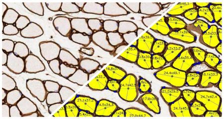
| Electrical impedance myography detects dystrophin-related muscle changes in mdx mice | Hiyoshi T, Zhao F, Baba R, Hirakawa T, Kuboki R, Suzuki K, Tomimatsu Y, O'Donnell P, Han S, Zach N, Nakashima M | 2023 | Research Square | Myology | Area Quantification, Muscle Fiber | HALO |
| Effects of the purified dry extract of fermented ginseng BST204 on muscle fiber regeneration | Jo S, Park Y, Chang Y, Moon J, Lee S, Lee H, Kim M, Kim D, Bae S, Park S, Yun H, You J, Im M, Han H, Kim S, Jin D | 2023 | Biochemistry and Biophysics Reports | Myology | Muscle Fiber | HALO |
| Rotator cuff muscle fibrosis can be assessed using ultrashort echo time magnetization transfer MRI with fat suppression | Chang EY, Suprana A, Tang Q, Cheng X, Fu E, Orozco E, Jerban S, Shah S, Du J, Ma Y | 2023 | NMR in Biomedicine | Fibrosis | Muscle Fiber | HALO |
| Heteroduplex oligonucleotide technology boosts oligonucleotide splice switching activity of morpholino oligomers in a Duchenne muscular dystrophy mouse model | Hasegawa J, Nagata T, Ihara K, Tanihata J, Ebihara S, Yoshida-Tanaka K, Yanagidaira M, Ohara M, Sasaki A, Nakayama M, Yamamoto S, Ishii T, Iwata-Hara R, Naito M, Miyata K, Sakaue F, Yokota T | 2024 | Nature Communications | Myology | Area Quantification, Muscle Fiber | HALO |
| Peripheral neural interfaces: Skeletal muscles are hyper-reinnervated according to the axonal capacity of the surgically rewired nerves | Tereshenko V, Dotzauer D C, Schmoll M, Harnoncourt L, Carrero Rojas G, Gfrerer L, Eberlin K R, Austen Jr. W G, Blumer R, Farina D, Aszmann O C | 2024 | Science Advances | Neuroscience | Muscle Fiber | HALO |
| Selpercatinib mitigates cancer cachexia independent of anti-tumor activity in the HT1080 tumor model | Skatri U, Gouda MA, Pandey S, Chauhan NK, Shen T, Hu X, Li M, Huang S, Subbiah V, Wu J | 2025 | Cancer Letters | Oncology | Muscle Fiber | HALO |