


HALO® Analysis Applications in Neuroscience Research
15 August 2018 | In this one hour webinar, Indica Labs’ Application Scientist, Alyssa Myers, will discuss how the HALO platform can be used for

Simultaneously analyze an unlimited number of fluorescent dyes and measure object density, area, diameter, and intensity, as well as colocalizations, if applicable.
Learn More
Quantify microglial activation in fluorescence based on detection of microglia, soma, and processes, by counting branch points, and by determining area, length, and thickness of processes.
Learn More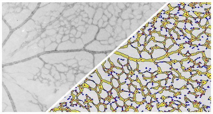
Quantify branch ends, area, length, and more for branched structures such as retinal vessels and cortical neurons in brightfield images.
Learn More