

HALO Image Analysis Masterclass Series: February-March 2021
Spring of 2021 | Indica Labs is excited to continue our HALO® Masterclass Webinar Series this winter. Each masterclass webinar will offer a deep dive

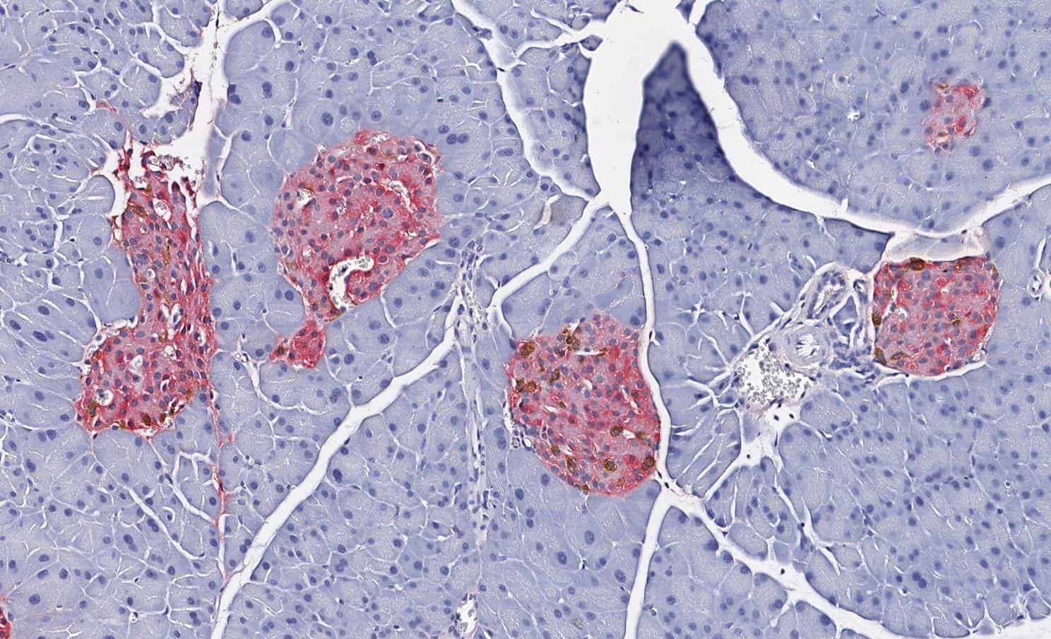
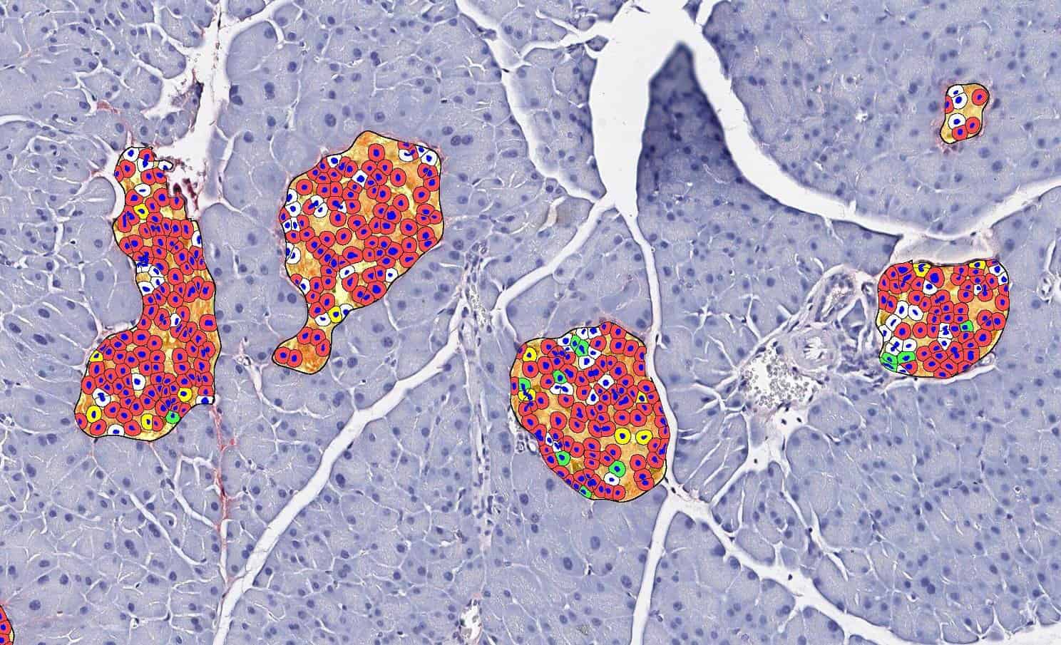
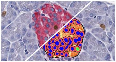
Quantify expression of up to five brightfield stains in any cellular compartment - membrane, nucleus or cytoplasm.
Learn More
Separate multiple tissue classes across a tissue using a learn-by-example approach. Can be used in conjunction with all other modules (fluorescent and brightfield) to select specific tissue classes for further analysis.
Learn More
Quantify the area, diameter, perimeter, and number of white spaces per region of interest in brightfield images. Ideally suited for analysis of lipids in brown and white adipose tissue, lipid droplets in liver tissue (steatosis), and alveoli area in lung.
Learn More