| Phase I trial of ADP-A2AFP TCR T-cell therapy in patients with advanced hepatocellular or gastric hepatoid carcinoma | Meyer T, Finn R S, Borad M, Mahipal A, Edeline J, Houot R, Hausner P F, Hollebecque A, Goyal L, Frigault M, Evans T R J, Wong K M, Tan B R, Mitry E, Sarker D, Feun L, El-Rayes B, Thistlethwaite F, Kaseb A, Alese O, Jin Z, Cirillo C, Bruix J, Roddie C, Noto P, Fayngerts S, Cristiani S, Sampson J, Bai J, Isabelle M, Broad R, Sun A, Norry E, Sangro B | 2025 | Journal of Hepatology | Immuno-oncology | Highplex FL, ISH-IHC | HALO |
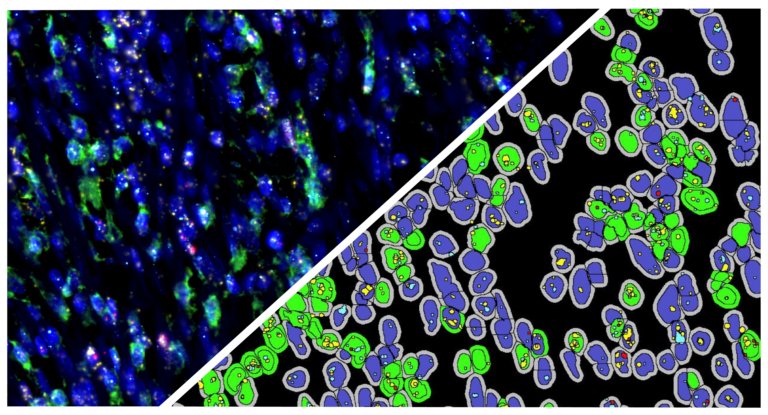
| Detection of engineered T cells in FFPE tissue by multiplex in situ hybridization and immunohistochemistry | Wright JH, Huang L-Y, Weaver S, Archila LD, McAfee MS, Hirayama AV, Chapuis AG, Bleakley M, Rongvaux A, Turtle CJ, Chanthaphavong S, Campbell JS, Pierce RH | 2020 | Journal of Immunological Methods | Immunology | ISH/FISH, ISH-IHC/FISH-IF | HALO |
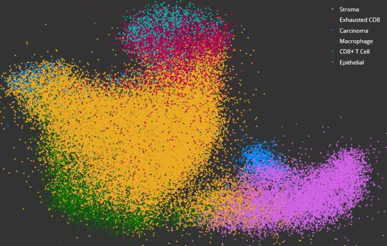
| Spatially organized multicellular immune hubs in human colorectal cancer | Pelka K, Hofree M, Chen J, Sarkizova S, Pirl J, Jorgji V, Bejnood A, Dionne D, Ge W, Xu K, Chao S, Zollinger D, Lieb D, Reeves J, Fuhrman C, Hoang M, Delorey T, Nguyen L, Waldman J, Klapholz M, Wakiro I, Cohen O, Albers, J, Smillie C, Cuoco M, Wu J, Su M, Yeung J, Vijaykumar B, Magnuson A, Asinovski N, Moll T, Goder-Reise M, Applebaum A, Brais L, DelloStritto L, Denning S, Phillips S, Hill E, Meehan J, Frederick D, Sharova T, Kanodia A, Todres E, Jane-Valbuena J, Biton M, Izar B, Lambden C, Clancy T, Bleday R, Melnitchouk N, Irani J, Kunitake H, Berger D, Srivastava A, Hornick J, Ogino S, Rotem A, Vigneau S, Johnson B, Corcoran R, Sharpe A, Kuchroo V, Ng K, Giannakis M, Nieman L, Boland G, Arguirre A, Anderson A, Rosenblatt-Rosen O, Regev A, Hachohen N | 2021 | Cell | Oncology | Spatial Analysis, ISH-IHC/FISH-IF | HALO |
| Detection of Epstein–Barr Virus in Periodontitis: A Review of Methodological Approaches | Tonoyan L, Chevalier M, Vincent-Bungnas S, Marsault R, Doglio A | 2021 | Mircroorganisms | Review | ISH/FISH, ISH-IHC/FISH-IF | HALO |
| Multiparameter immunohistochemistry analysis of HIV DNA, RNA and immune checkpoints in lymph node tissue | Richardson Z, Deleage C, Tutuka C, Walkiewica M, Del Rio-Estrada P, Pascoe R, Evans V, Reyesteran G, Gonzales M, Roberts-Thomson S, Gonzalez-Navarro M, Torres-Ruiz, F, Estes J, Lewin S, Cameron P | 2021 | Journal of Immunological Methods | Infectious Disease | ISH-IHC/FISH-IF | HALO |
| IL-1β Promotes Expansion of IL-33+ Lung Epithelial Stem Cells Following RSV Infection During Infancy | Vu L, Phan A, Hijano D, Siefker D, Tillman H, Cormierr S | 2021 | American Journal of Respiratory Cell and Molecular Biology | Infectious Disease | ISH-IHC | HALO |
| Satellite repeat RNA expression in epithelial ovarian cancer associates with a tumor immunosuppressive phenotype | Porter R, Sun S, Flores M, Berzolla E, You E, Phillips I, KC N, Desai N, Tai E, Szabolcs A, Lang E, Pankaj A, Raabe M, Thapar V, Xu K, Nieman L, Rabe D, Kolin D, Stover E, Pepin D, Stott S, Deshpande V, Liu J, Solovyov A, Matulonis U, Greenbaum B, Ting D | 2022 | The Journal of Clinical Investigation | Immuno-oncology | ISH/FISH, ISH-IHC/FISH-IF | HALO |
| Annexin A2/TLR2/MYD88 pathway induces arginase 1 expression in tumor-associated neutrophils | Zhang H, Zhu X, Friesen T, Kwak J, Pisarenko T, Mekvanich S, Velasco M, Randolph T, Kargl J, Houghton A | 2022 | Journal of Clinical Investigation | Immuno-oncology | ISH-IHC/FISH-IF | HALO |
| Spatiotemporal transcriptome analysis reveals critical roles for mechano-sensing genes at the border zone in remodeling after myocardial infarction | Yamada S, Ko T, Hatsuse S, Nomura S, Zhang B, Dai Z, Inoue S, Kubota M, Sawami K, Yamada T, Sassa T, Katagiri M, Fujita K, Katoh M, Ito M, Harada M, Toko H, Takeda N, Morita H, Aburatani H, Komuro I | 2022 | Nature Cardiovascular Research | Myology | ISH-IHC/FISH-IF | HALO |
| Loss of functional System x-c uncouples aberrant postnatal neurogenesis from epileptogenesis in the hippocampus of Kcna1-KO mice | Aloi M, Thompson S, Quartapella N, Noebels J | 2022 | Cell Reports | Neuroscience | ISH-IHC/FISH-IF | HALO |
| The potassium channel auxiliary subunit Kvβ2 (Kcnab2) regulates Kv1 channels and dopamine neuron firing | Yee J, Rastani A, Soden M | 2022 | Journal of Neurophysiology | Neuroscience | ISH-IHC/FISH-IF | HALO |
| Transcriptional vulnerabilities of striatal neurons in human and rodent models of Huntington’s disease | Matsushima A, Pineda S, Crittenden J, Lee H, Galani K, Mantero J, Tombaugh G, Kellis M, Heiman M, Graybiel A | 2023 | Nature Communications | Neuroscience | Area Quantification, ISH-IHC/FISH-IF | HALO |