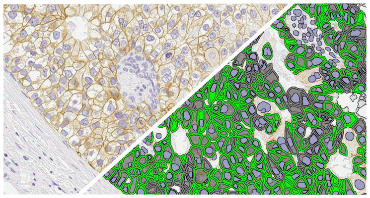
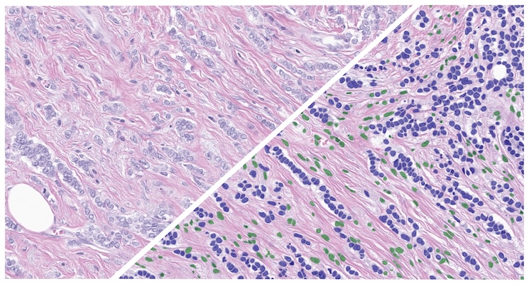
New! ADC Target Scoring Module
Quantify expression of an unlimited number of biomarkers in any cellular compartment – membrane, nucleus or cytoplasm.
New! ADC Target Scoring Module Read More »
Quantify expression of an unlimited number of biomarkers in any cellular compartment – membrane, nucleus or cytoplasm.
New! ADC Target Scoring Module Read More »
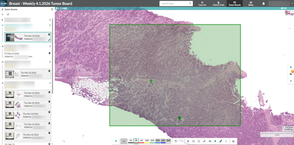
HALO AP® AI Powered, Pathologist Driven The CE-IVDR marked, AI-powered image management system built to digitize clinical pathology workflows and allow pathologists to work anytime,
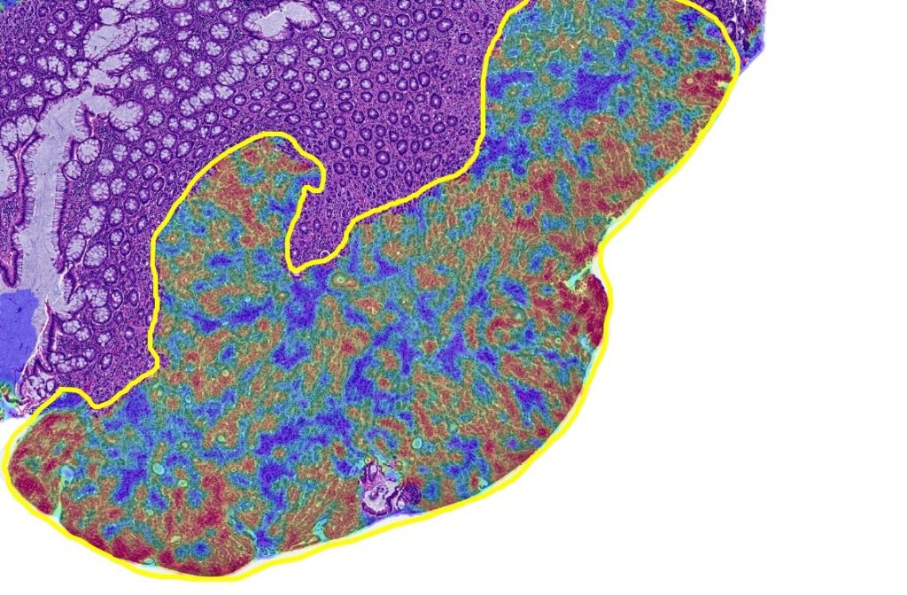
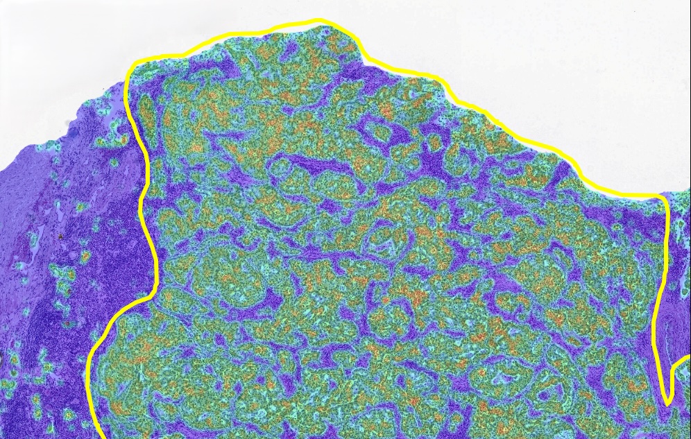
Reliable tumor content analysis for colorectal carcinoma specimens.
HALO Macrodissection Solutions Read More »
AI for Prostate Tumor Detection and Gleason Grading Prostate AI is an AI-powered digital pathology analysis tool designed to identify and classify prostate adenocarcinoma in
The Breast H&E Cancer Cell Phenotyper App is a pre-trained HALO AI phenotyper designed to detect and quantify cancer cells across whole slide H&E-stained images of breast cancer tissue.
Breast H&E Cancer Cell Phenotyper Read More »
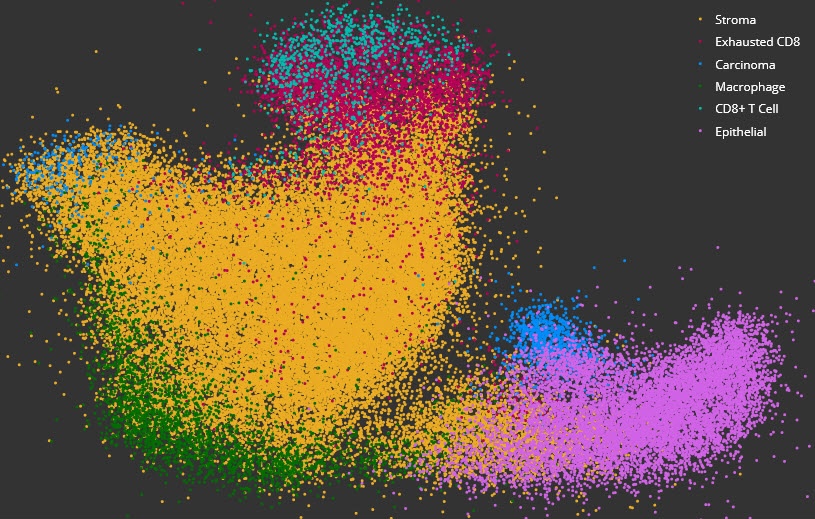
Acquire deeper insights into complex data sets using dimensional reduction and unsupervised clustering with interactive plotting.
High Dimensional Analysis Module Read More »
Reliable tumor content analysis for colorectal carcinoma specimens.
CRC Macrodissect AI Read More »
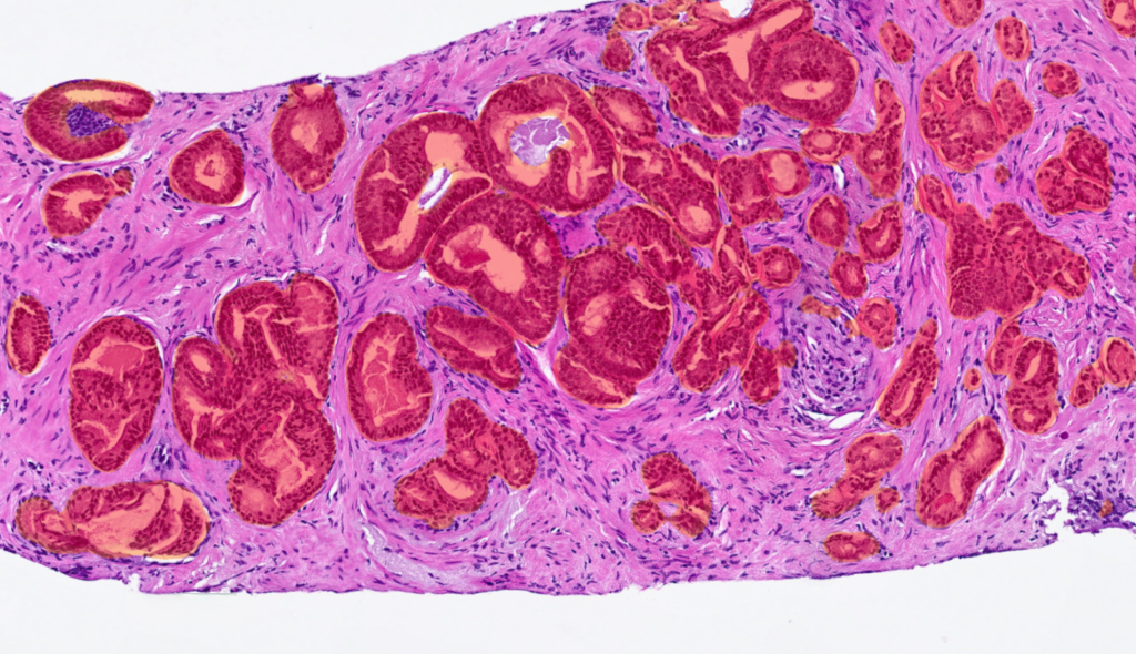
Reliable tumor content analysis for non-small cell lung cancer specimens.
Lung Macrodissect AI Read More »

The Non-Small Cell Lung Cancer (NSCLC) H&E Tumor Tissue Detection App is a pre-trained HALO AI masking classifier designed to segment tumor, stroma, necrosis/other, and glass area across H&E-stained whole slide images of NSCLC.
NSCLC H&E Tumor Tissue Detection Read More »

The Ovarian H&E Tumor Tissue Detection App is a pre-trained HALO AI masking classifier designed to segment tumor, stroma, necrosis/other, and glass area across H&E-stained whole slide images of ovarian cancer.
Ovarian H&E Tumor Tissue Detection Read More »