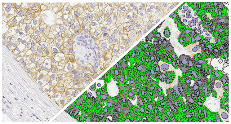
New! ADC Target Scoring Module
Quantify expression of an unlimited number of biomarkers in any cellular compartment – membrane, nucleus or cytoplasm.
New! ADC Target Scoring Module Read More »
Quantify expression of an unlimited number of biomarkers in any cellular compartment – membrane, nucleus or cytoplasm.
New! ADC Target Scoring Module Read More »
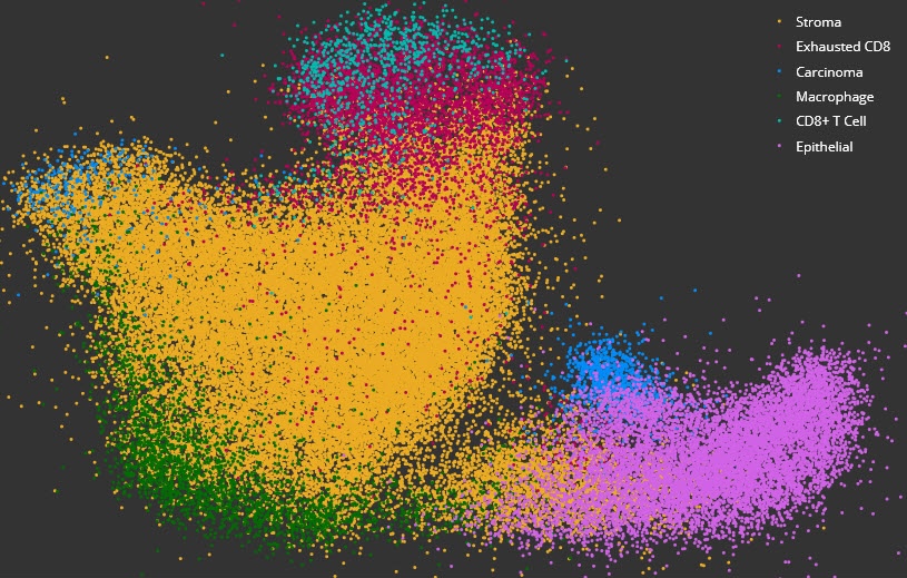
Acquire deeper insights into complex data sets using dimensional reduction and unsupervised clustering with interactive plotting.
High Dimensional Analysis Module Read More »

Quantify branch ends, area, length, and more for branched structures such as retinal vessels and cortical neurons in fluorescent images.
Branch Structure FL Read More »

Quantify microglial activation in fluorescence based on detection of microglia, soma, and processes, by counting branch points, and by determining area, length, and thickness of processes.
Microglial Activation FL Read More »
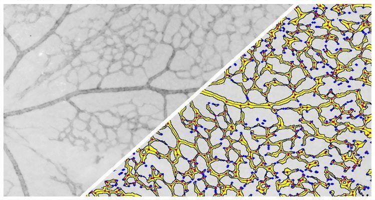
Quantify branch ends, area, length, and more for branched structures such as retinal vessels and cortical neurons in brightfield images.
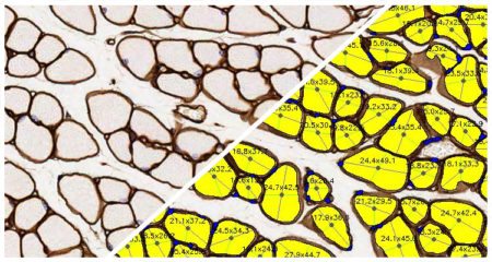
Quantify area, diameter, and perimeter of muscle fibers stained with laminin or other fiber membrane stains.

Process multiple images in parallel to maximize image analysis throughput and minimize analysis job turnaround times.
Analysis Cluster Add-on Read More »

Count and measure pancreatic islets in fluorescent images and quantify the islet area and cells positive for up to three islet-specific dyes.

Simultaneously analyze up to five chromogenic stains and measure object density, area, diameter, and optical density, as well as colocalizations, if applicable.
Object Colocalization Read More »

Quantify expression of an unlimited number of biomarkers in any cellular compartment – membrane, nucleus or cytoplasm.