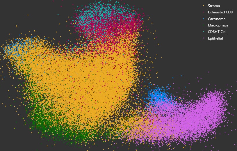
High Dimensional Analysis Module
Acquire deeper insights into complex data sets using dimensional reduction and unsupervised clustering with interactive plotting.
High Dimensional Analysis Module Read More »
Acquire deeper insights into complex data sets using dimensional reduction and unsupervised clustering with interactive plotting.
High Dimensional Analysis Module Read More »

Simultaneously analyze up to five chromogenic stains and measure object density, area, diameter, and optical density, as well as colocalizations, if applicable.
Object Colocalization Read More »

Quantify expression of an unlimited number of biomarkers in any cellular compartment – membrane, nucleus or cytoplasm.

The TMA Add-on provides a productivity-enhancing workflow for tissue microarray analysis by enabling automated, high-throughput segmentation and batch analysis of whole slide TMA images.
TMA (Tissue Microarray) Add-On Read More »

Simultaneously analyze an unlimited number of fluorescent dyes and measure object density, area, diameter, and intensity, as well as colocalizations, if applicable.
Object Colocalization FL Read More »

Plot cells and objects from one or more images and perform nearest neighbor analysis, proximity analysis, and tumor infiltration analysis.
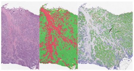
Analyze serial tissue sections stained with different markers or a single tissue section which has been stained, stripped, and re-stained for multiple markers.
Serial Section Analysis Add-on Read More »
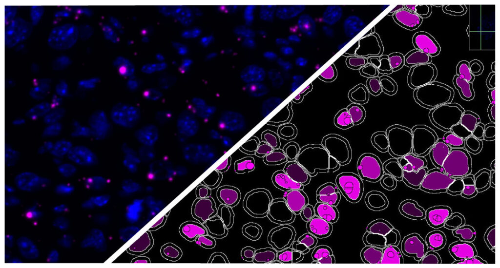
Simultaneously analyze an unlimited number of fluorescent nucleic acid probes on a cell-by-cell basis, measuring spot numbers and area per cell and compartment, and calculated H-scores for each probe.

Simultaneously analyze up to three chromogenic and/or silver-labelled DNA or RNA ISH probes on a cell-by-cell basis, measuring spot numbers and area per cell and compartment, and calculated H-scores for each probe.

Use the HALO® FISH-IF module and reagents from Molecular Instruments or ACD, a Bio-techne brand, to simultaneously analyze an unlimited number of fluorescently-labeled DNA/RNA ISH probes and immunofluorescent protein biomarkers on a cell-by-cell basis.