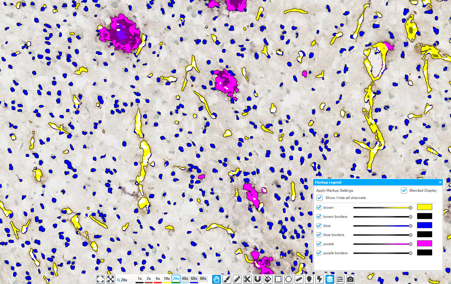
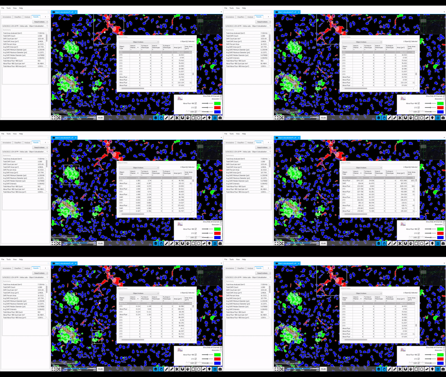
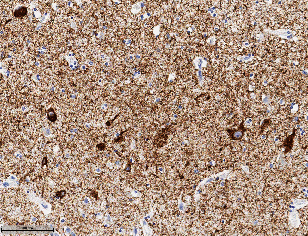
Simultaneously analyze up to five chromogenic stains with the HALO® Object Colocalization module. This module is ideally suited for neuroscience applications that require counting and measuring multiple object types, such as amyloid plaques, neurofibrillary tangles, or microglia. Choose to output data on a per-object level for in-depth analysis of object density, area, diameter, and optical density, as well as colocalizations, if applicable. After analysis, leverage interactive markups to dynamically explore objects and colocalization.
Try it out! Click here to initiate your free proof-of-concept HALO image analysis.
File formats supported by the HALO image analysis platform:
- Non-proprietary (JPG, TIF, OME.TIFF)
- Nikon (ND2)
- 3D Histech (MRXS)
- Akoya (QPTIFF, component TIFF)
- Olympus / Evident (VSI)
- Hamamatsu (NDPI, NDPIS)
- Aperio (SVS, AFI)
- Zeiss (CZI)
- Leica (SCN, LIF)
- Ventana (BIF)
- Philips (iSyntax, i2Syntax)
- KFBIO (KFB, KFBF)
- DICOM (DCM*)
*whole-slide images

Announcing HALO® Vacuole, Object Colocalization BF, and Object Colocalization FL Modules Supporting Interactive Markups
We’re excited to announce the launch of the newly updated Vacuole, Object Colocalization BF, and Object Colocalization FL modules supporting interactive markups.

Interactive Markup Images: Providing a Dynamic Look at HALO® Analysis Results
At Indica Labs, our HALO platforms are optimized for ease-of-use as well as powerful, accurate analysis, and include multiple features to help users get the

Announcing the Object Colocalization 2.1 modules for BF and FL
In this blog post, you can learn about some of the new features in these modules, where to find the user guides, tutorial videos, and

Masterclass Webinar: Neurobiology Image Analysis with HALO and HALO AI
29 September 2022 | Join us for this 1-hour webinar for a live demonstration of neurobiology image analysis using HALO® and HALO AI. Dr. Levi

Maximizing use of HALO® and HALO AI for a comprehensive image analysis for HUMAN Brain FFPE Tissue samples in Alzheimer’s Disease
25 May 2022 | In this 60-min webinar, Learn how HALO and HALO AI are advancing neuropathology research at the UW Medicine Biorepository and Integrated

Webinar | HALO® Image Analysis Masterclass Series: Object-based Analysis
28 January 2021 | 8:00 AM – 9:00 AM PST | 11:00 AM – 12:00 PM EST | 4:00 PM – 5:00 PM GMT |

HALO® Analysis Applications in Neuroscience Research
15 August 2018 | In this one hour webinar, Indica Labs’ Application Scientist, Alyssa Myers, will discuss how the HALO platform can be used for
Publication Spotlight
The table below includes publications that cite the Object Colocalization module.
Your publication not on the list? Drop us an email to let us know about it!
| Title | Authors | Year | Journal | Application | HALO Modules | Product |
|---|---|---|---|---|---|---|
| Photoacoustic imaging biomarkers for monitoring biophysical changes during nanobubble-mediated radiation treatment | Hysi E, Fadhel MN, Wang Y, Sebastian JA, Giles A, Czarnota GJ, Exner AA, Kolios MC | 2020 | Photoacoustics | Other | Classifier, Area Quantification, Object Colocalization | HALO |
| Automated macrophage counting in DLBCL tissue samples: a ROF filter based approach | Wagner M, Hansel R, Reinke S, Richter J, Altenbuchinger M, Braumann U-D, Spang R, Loffler Mk Klapper W | 2019 | Biological Proectures Online | Oncology | Object Colocalization | HALO |
| Relationship between mismatch repair immunophenotype and long-term survival in patients with resected periampullary adenocarcinoma | Heby M, Lundgren S, Nodin B, Elebro J, Eberhard J, JirstrÀÜm K | 2018 | Journal of Translational Medicine | Oncology, Immuno-oncology | Object Colocalization | HALO |
| Age-related pathology after adenoviral overexpression of the leucine-rich repeat kinase 2 in the mouse striatum | Kritzingerab A, Ferger B, Gillardon F, Stierstorfer B, Birk G, Kochanek S, Ciossek T | 2018 | Neurobiology of Aging | Neuroscience | Area Quantification, Object Colocalization, Microglia | HALO |
| Pharmacogenetic neuronal stimulation increases human tau pathology and trans-synaptic spread of tau to distal brain regions in mice | Schultz MK, Gentzel R, Usenovic M, Gretzul C, Ware C, Parmentier-Batteur S, Schachter JB, Zariwala HA | 2018 | Neurobiology of Disease | Neuroscience | Object Colocalization | HALO |
| Comparison of Biologic Effect and Particulate Embolization after Femoral Artery Treatment with Three Drug-Coated Balloons in Healthy Swine Model | Torii S, Jinnouchi H, Sakamoto A, Romero ME, Kolodgie FD, Virmani R, Finn AV | 2018 | Journal of Vascular and Interventional Radiology | Other | Object Colocalization | HALO |
| Aging African green monkeys manifest transcriptional, pathological, and cognitive hallmarks of human Alzheimer's disease | Cramer PE, Gentzel RC, Tanis KQ, Vardigan J, Wang Y, Connolly B, Manfre P, Lodge K, Renger JJ, Zerbinatti C, Uslaner JM | 2017 | Neurobiology of Aging | Neuroscience | Object Colocalization | HALO |
| Chronic Verubecestat Treatment Suppresses Amyloid Accumulation in Advanced Aged Tg2576-A?PPswe Mice Without Inducing Microhemorrhage | Villarreal S, Zhao F, Hyde LA, Holder D, Forest T, Sondey M, Chen X, Sur C, Parker EM, Kennedy ME | 2017 | Journal of Alzheime's Disease | Neuroscience | Object Colocalization | HALO |
| The integrative clinical impact of tumor-infiltrating T lymphocytes and NK cells in relation to B lymphocyte and plasma cell density in esophageal and gastric adenocarcinoma | Svensson MC, Warfvinge CF, Fristedt R, Hedner C, Borg D, Eberhard J, Micke P, Nodin B, Leandersson K, Jirstrom K | 2017 | Oncotarget | Immuno-oncology | Object Colocalization | HALO |
| Resistance to Anti-VEGF Therapy Mediated by Autocrine IL6/STAT3 Signaling and Overcome by IL6 Blockade | Eichten A, Su J, Adler AP, Zhang L, Ioffe E, Parveen AA, Yancopoulos GD, Rudge J, Lowy I, Lin HC, MacDonald D, Daly C, Duan X, Thurston G | 2016 | Cancer Research | Oncology, Immuno-oncology | Cytonuclear, Object Colocalization | HALO |
| Cerebral Protection During MitraClip Implantation: Initial Experience at 2 Centers | Frerker C, Schl¸ter M, Sanchez OD, Reith S, Romero ME, Ladich E, Schrˆder J, Schmidt T, Kreidel F, Joner M | 2016 | JACC: Cardiovascular Interventions | Myology | Object Colocalization | HALO |
| The Prognostic Impact of NK/NKT Cell Density in Periampullary Adenocarcinoma Differs by Morphological Type and Adjuvant Treatment | Lundgren S, Warfvinge CF, Elebro J, Heby M, Nodin B, Krzyzanowska A, Bjartell A, Leandersson K, Eberhard J, Jirstrom K | 2016 | PLOS ONE | Oncology, Immuno-oncology | Object Colocalization | HALO |
| Semi-Automated Digital Image Analysis of Pick's Disease and TDP-43 Proteinopathy | Irwin DJ, Byrne MD, McMillan CT, Cooper F, Arnold SE, Lee EB, Van Deerlin VM, Xie SX, Lee VM, Grossman M, Trojanowski JQ | 2015 | Journal of Histochemistry & Cytochemistry | Neuroscience | Object Colocalization, Layer Thickness | HALO |
| Engineered red blood cells as an off-the-shelf allogeneic anti-tumor therapeutic | Zhang X, Lou M, Dastagir SR, Nixon M, Khamhoung A, Schmidt A, Lee A, Subbiah N, McLaughlin DC, Moore CL, Gribble M, Bayhi N, Amin V, Pepi R, Pawar S, Lyford TJ, Soman V, Mellen J, Carpenter CL, Turka LA, Wickham Tj, Chen TF | 2021 | Nature Communications | Oncology | Object Colocalization, Highplex FL | HALO |
| Wound healing with topical BRAF inhibitor therapy in a diabetic model suggests tissue regenerative effects | Escuin-Ordinas H, Liu Y, Hufo W, Dimatteo R, Huang R, Krystofinski P, Azhdam A, Lee J, Comin-Anduix B, Cochran A, Lo R, Segura T, Scumpia P, Ribas A | 2021 | PLOS ONE | Dermatology | Object Colocalization, Multiplex IHC | HALO |
| Acute brain inflammation, white matter oxidative stress, and myelin deficiency in a model of neonatal intraventricular hemorrhage | Goulding DS, Vogel RC, Gensel JC, Morganti JM, Stromberg AJ, Miller BA | 2020 | Journal of Neuroscience | Immunology, Neuroscience | Area Quantification, Object Colocalization | HALO |
| Promyelocytic leukemia protein promotes the phenotypic switch of smooth muscle cells in atherosclerotic plaques of human coronary arteries | Karle W, Becker S, Stenzel P, Knosalla C, Siegel G, Baum O, Zakrzewicz A, Berkholz J | 2021 | Clinical Science | Myology | Area Quantification, Object Colocalization | HALO |
| Mitochondria exert age-divergent effects on recovery from spinal cord injury | Stewart A, McFarlane K, Vekaria H, Bailey W, Slone S, Tranthem L, Zhang B, Patel S, Sullivan P, Gensel J | 2021 | Experimental Neurology | Neuroscience | Object Colocalization | HALO |
| The GBA1 D409V mutation exacerbates synuclein pathology to differing extents in two alpha-synuclein models | Polinski N, Martinez T, Ramboz S, Sasner M, Herberth M, Switzer R, Ahmad S, Pelligrino L, Clark S, Marcus J, Smith S, Dave K, Frasier M | 2022 | Disease Models & Mechanisms | Neuroscience | Object Colocalization | HALO |
| Intraoperative abobotulinumtoxinA alleviates pain after surgery and improves general wellness in a translational animal model | Cornet S, Carre D, Limana L, Castel D, Meilin S, Horn R, Pons L, Evans S, Lezmi S, Kalinichev M | 2022 | Research Square | Other | Area Quantification, Object Colocalization | HALO |
| Combined Blockade of GARP:TGF-?1 and PD-1 Increases Infiltration of T Cells and Density of Pericyte-Covered GARP+†Blood Vessels in Mouse MC38 Tumors | Bertrand C, Van Meerbeeck P, de Streel G, Vaherto-Bleeckx N, Benhaddi F, Rouaud L, Noel A, Coulie P, van Baren N, Lucas S | 2022 | Frontiers of Immunology | Immuno-oncology | Cytonuclear, Object Colocalization | HALO |
| Inflammatory Pathways Are Impaired in Alzheimer Disease and Differentially Associated With Apolipoprotein E Status | Kloske C, Dugan A, Weekman E, Winder Z, Patel E, Nelson P, Fardo D, Wilcock D | 2021 | Journal of Neuropathology & Experimental Neurology | Neuroscience | Object Colocalization | HALO |
| Aristolochic acid I promoted clonal expansion but did not induce hepatocellular carcinoma in adult rats | Liu Y, Lu H, Qi X, Xing G, Wang X, Yu P, Liu L, Yang F, Ding X, Deng Z, Gong L, Ren J | 2021 | Acta Pharmacologica Sinica | Oncology | Object Colocalization | HALO |
| High endothelial venules associated with better prognosis in esophageal squamous cell carcinoma | Li H, Tang L, Han X, Zhong L, Gao W, Chen Y, Huang J, Wen Z | 2022 | Annals of Diagnostic Pathology | Oncology | Object Colocalization, Spatial Analysis, Highplex FL | HALO |
| Somatostatin slows Aβ plaque deposition in aged APPNL-F/NL-F mice by blocking Aβ aggregation | Williams D, Yan B, Wang H, Negm L, Sackmann C, Verkuyl C, Rezai-Stevens V, Eid S, Vediya N, Sato C, Watts J, Wille H, Schmitt-Ulms G | 2023 | Scientific Reports | Neuroscience | Object Colocalization | HALO |
| The localization of molecularly distinct microglia populations to Alzheimer's disease pathologies using QUIVER | Shahidehpour R, Nelson A, Sanders L, Embry C, Nelson P, Bachstetter A | 2023 | Acta Neuropathologica Communications | Neuroscience | Area Quantification, Object Colocalization, Spatial Analysis, Registration | |
| Soluble TNF mediates amyloid-independent, diet-induced alterations to immune and neuronal functions in an Alzheimer’s disease mouse model | MacPherson K, Eidson L, Houser M, Weiss B, Gollihue J, Herrick M, Rodrigues M, Sniffen L, Weekman E, Hamilton A, Kelly S, Oliver D, Yang Y, Chang J, Sampson T, Norris C, Tansey M | 2023 | Frontiers in Cellular Neuroscience | Neuroscience | Object Colocalization | HALO |
| Chronic immune activation and gut barrier dysfunction is associated with neuroinflammation in ART-suppressed SIV+ rhesus macaques | Byrnes S, Busman-Sahay K, Angelovich T, Younger S, Taylor-Brill S, Nekorchuk M, Bondoc S, Dannay R, Terry M, Cochrane C, Jenkins T, Roche M, Deleage C, Bosinger S, Paiardini M, Brew B, Estes J, Churchill M | 2023 | PLOS Pathogens | Infectious Disease | Area Quantification, Object Colocalization, ISH, Highplex FL | HALO |
| Early chronic suppression of microglial p38α in a model of Alzheimer’s disease does not significantly alter amyloid-associated neuropathology | Braun D, Frazier H, Davis V, Coleman M, Rogers C, Van Eldik L | 2023 | PLOS One | Neuroscience | Object Colocalization | HALO |
| Modifying antibody-FcRn interactions to increase the transport of antibodies through the blood-brain barrier | Tien J, Leonoudakis D, Petrova R, Trinh V, Taura T, Sengupta D, Jo L, Sho A, Yun Y, Doan E, Jamin A, Hallak H, Wilson D, Stratton J | 2023 | mABS | Neuroscience | Classifier, Area Quantification, Object Colocalization | HALO |
| Atorvastatin rescues hyperhomocysteinemia-induced cognitive deficits and neuroinflammatory gene changes | Weekman E, Johnson S, Rogers C, Sudduth T, Xie K, Qiao Q, Fardo D, Bottiglieri T, Wilcock D | 2023 | Journal of Neuroinflammation | Neuroscience | Object Colocalization | HALO |
| Altered expression, but small contribution, of the histone demethylase KDM6A in obstructive uropathy in mice | Hong LYQ, Yeung ESH, Tran DT, Yerra VGY, Kaur HK, Kabir MDG, Advani SL, Liu Y, Batchu SN, Advani A | 2023 | Disease Models & Mechanisms | Gastroenterology | Area Quantification, Object Colocalization | HALO |
| LINC01638 Sustains Human Mesenchymal Stem Cell Self-Renewal and Competency for Osteogenic Cell Fate | Gordon J, Tye C, Banerjee B, Ghule P, Wijnen A, Kabala F, Page N, Falcone M, Stein J, Stein G, Lian J | 2023 | Research Square | Other | Object Colocalization, FISH | HALO |
| Distinct Patterns of Hippocampal Pathology in Alzheimer's Disease with Transactive Response DNA-binding Protein 43 | Minogue G, Kawles A, Zouridakis A, Keszycki R, Macomber A, Lubbat V, Gill N, Mao Q, Flanagan ME, Zhang H, Castellani R, Bigio EH, Mesulam M-M, Geula C, Gefen T | 2023 | Annals of Neurology | Neuroscience | Object Colocalization | HALO |
| Regional and cellular iron deposition patterns predict clinical subtypes of multiple system atrophy | Lee S, Martinez-Valbuena I, Lang AE, Kovacs GG | 2023 | Research Square | Neuroscience | Area Quantification, Object Colocalization | HALO |
| Empowering Renal Cancer Management with AI and Digital Pathology: Pathology, Diagnostics and Prognosis | Ivanova E, Fayzullin A, Grinin V, Ermilov D, Arutyunyan A, Timashev P, Shekhter A | 2023 | Biomedicines | Review | Classifier, Area Quantification, Object Colocalization | HALO, HALO AI |
| Association between APOE genotype and microglial cell morphology | Kloske C, Gearon M, Weekman E, Rogers C, Patel E, Bachstetter A, Nelson P, Wilcock D | 2023 | Journal of Neuropathology & Experimental Neurology | neuroscience | Object Colocalization, Microglia | HALO |
| Disease pathology signatures in a mouse model of Mucopolysaccharidosis type IIIB | Petrova R, Patil AR, Trinh V, McElroy KE, Bhakta M, Tien J, Wilson DS, Warren L, Stratton JR | 2023 | Scientific Reports | Neuroscience, Metabolism | Classifier, Area Quantification, Object Colocalization | HALO |
| Distinct Involvement of the Cranial and Spinal Nerves in Progressive Supranuclear Palsy | Tanaka H, Martinez-Valbuena I, Forrest SL, Couto B, Reyes NG, Morales-Rivero A, Lee S, Li J, Karakani AM, Tang-Wai DF, Tator C, Khadadadi M, Sadia N, Tartaglia MC, Lang AE, Kovacs GG | 2023 | Brain | Neuroscience | Object Colocalization | HALO |
| A proteome-wide screen identifies the calcium binding proteins, S100A8/S100A9, as clinically relevant therapeutic targets in aortic dissection | Jiang H, Zhao Y, Su M, Sun L, Chen M, Zhang Z, Ilyas I, Wang Z, Little P J, Wang L, Weng J, Ge J, Xu S | 2023 | Pharmacological Research | Other | Object Colocalization | HALO |
| ApoER2-Dab1 disruption as the origin of pTau-associated neurodegeneration in sporadic Alzheimer’s disease | Ramsden C, Zamora D, Horowitz M, Jahanipour J, Calzada E, Li X, Keyes G, Murray H, Curtis M, Faull R, Sedlock A, Maric D | 2023 | Acta Neuropathologica Communications | Neuroscience | Area Quantification, Object Colocalization | HALO |
| Induction of double-strand breaks with the non-steroidal androgen receptor ligand flutamide in patients on androgen suppression: a study protocol for a randomized, double-blind prospective trial | Lee E, Coulter J, Mishra A, Caramella-Pereira F, Demarzo A, Rudek M, Hu C, Han M, DeWeese TL, Yegnasubramanian S, Song DY | 2023 | Trials | Oncology | Object Colocalization, ISH | HALO |
| A microtubule stabilizer ameliorates protein pathogenesis and neurodegeneration in mouse models of repetitive traumatic brain injury | Zhao X, Zeng W, Xu H, Sun Z, Hu Y, Peng B, McBride JD, Duan J, Deng J, Zhang B, Kim SJ, Zoll B, Saito T, Sasaguri H, Saido TC, Ballatore C, Yao H, Wang Z, Trojanowski JQ, Brunden KR, Lee VMY, He Z | 2023 | Science Translational Medicine | neuroscience | Area Quantification, Object Colocalization, Multiplex IHC | HALO |
| Amelioration of Tumor-Promoting Microenvironment via Vascular Remodeling and CAF Suppression using E7130: Biomarker Analysis by Multi-modal Imaging Modalities | Ito K, Yamaguchi M, Semba T, Tabata K, Tamura M, Aoyama M, Abe T, Asano O, Terada Y, Funashashi Y, Fujii H | 2023 | Molecular Cancer Therapeutics | Oncology | Area Quantification, Object Colocalization | HALO |
| Retinoic acid signaling regulates spatiotemporal specification of human green and red cones | Hadyniak SE, Hagen JF, Eldred KC, Brenerman B, Hussey KA, McCoy RC, Sauria MEG, Kuchenbecker JA, Reh T, Glass I, Neitz M, Neitz J, Taylor J, Johnston Jr RJ | 2024 | Plos Biology | Other | Object Colocalization | HALO |
| Female mice display sex-specific differences in cerebrovascular function and subarachnoid haemorrhage-induced injury | Dinh DD, Wan H, Lidington D, Bolz SS | 2024 | EBioMedicine | Neuroscience | Object Colocalization, Microglia | HALO |
| Association between APOE genotype and microglial cell morphology | Kloskey CM, Gearon MD, Weekman EM, Rogers C, Patel E, Bachstetter A, Nelson PT, Wilcock DM | 2023 | Science Advances | Neuroscience | Object Colocalization, Microglia | HALO |
Related HALO Modules
Plot cells and objects from one or more images and perform nearest neighbor analysis, proximity analysis, and tumor infiltration analysis.
Learn MoreSeparate multiple tissue classes across a tissue using a learn-by-example approach. Can be used in conjunction with all other modules (fluorescent and brightfield) to select specific tissue classes for further analysis.
Learn MoreDeconvolve up to five colors in brightfield and measure positive area and average optical density for each stain and stain colocalization (where applicable).
Learn MoreUse the arrows above to view additional related modules
Want to Learn More?
Fill out the form below to request information about any of our software products.
You can also drop us an email at info@indicalab.com
Products & Services
Interested in purchasing or learning more about our products and services? Our highly trained application scientists are a couple of clicks away.
Software Maintenance & Support Coverage
Interested in purchasing an SMS plan? We would be happy to give you a quote.
Technical Support
Need technical support? Our IT specialists are here to help.


