Muscle Fiber
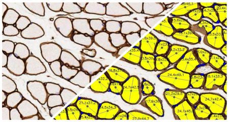
Quantify area, diameter, and perimeter of muscle fibers stained with laminin or other fiber membrane stains.
Quantify area, diameter, and perimeter of muscle fibers stained with laminin or other fiber membrane stains.

Simultaneously analyze up to five chromogenic stains and measure object density, area, diameter, and optical density, as well as colocalizations, if applicable.
Object Colocalization Read More »

Quantify fiber or membrane positivity for an unlimited number of fluorescent dyes along with fiber diameter, perimeter, and area.

Chris McKinnon, et al, Acta Neuropathologica Communications, 2020.
In this study, McKinnon et al identify overexpression of α-synuclein leading to catalytic impairment of the 26S proteosome in defined regions of rat brains. Brain tissue was fixed following α-synuclein overexpression for immunofluorescence studies of dopaminergic neurons which were quantified by the HALO image analysis platform. Future research will focus on characterizing the relationship between proteasome impairment and neurodegeneration.