Muscle Fiber
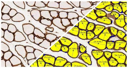
Quantify area, diameter, and perimeter of muscle fibers stained with laminin or other fiber membrane stains.
Quantify area, diameter, and perimeter of muscle fibers stained with laminin or other fiber membrane stains.

Simultaneously analyze up to five chromogenic stains and measure object density, area, diameter, and optical density, as well as colocalizations, if applicable.
Object Colocalization Read More »

Quantify microglial activation based on length and thickness of microglial processes.
Microglial Activation Read More »

Quantify fiber or membrane positivity for an unlimited number of fluorescent dyes along with fiber diameter, perimeter, and area.
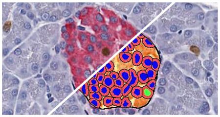
Count and measure pancreatic islets in brightfield images and quantify the islet area and cells positive for up to two islet-specific stains.
Islet Quantification Read More »