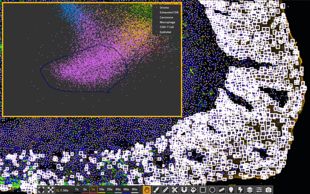
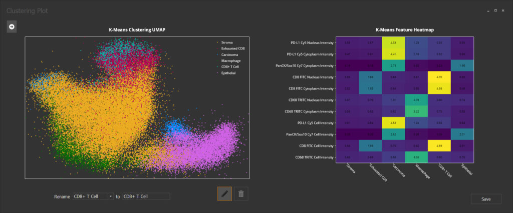
Multiplex IHC

Prognostic Value of CD8+ Lymphocytes in Hepatocellular Carcinoma and Perineoplastic Parenchyma Assessed by Interface Density Profiles in Liver Resection Samples

This study examined CD8+ cell distribution in hepatocellular carcinoma (HCC) and peritumoral liver tissue to investigate the overall survival (OS) and recurrence-free survival (RFS). It

Prognostic Value of CD8+ Lymphocytes in Hepatocellular Carcinoma and Perineoplastic Parenchyma Assessed by Interface Density Profiles in Liver Resection Samples Read More »

Spatial Biology: Revolutionizing Tissue Imaging and Analysis Using Spatial profiling

23 September 2021 | DigiBase invites you to attend a Spatial Biology webinar symposium to learn from experts in the field on how digital pathology, high-dimensional multiplexing, spatial and AI analytic tools, and spatial transcriptomic technologies are advancing tissue imaging and analysis.

Spatial Biology: Revolutionizing Tissue Imaging and Analysis Using Spatial profiling Read More »

Neoadjuvant immunotherapy leads to pathological responses in MMR-proficient and MMR-deficient early-stage colon cancers

Myriam Chalabi, et al, Nature Medicine, 2020.
Immunotherapy is known to be effective in late-stage, mismatch repair (MMR) deficient colorectal cancers but not in MMR proficient cancer. This study reports the early results from the NICHE clinical trial that is evaluating treatment of early stage, nonmetastatic preoperative colon cancer with a CTLA-4 inhibitor and a PD-1 inhibitor. Chalabi and colleagues report that the treatment was well tolerated and that a major pathological response was seen in 19/20 patients with MMR deficient tumors and 3/15 MMR proficient tumors. HALO image analysis software was used in this publication for analysis of T-cell biomarkers CD3, CD8, and FOXP3 on chromogenically stained tumor biopsies. First, image registration with HALO was performed of three serial tissue sections. Tissue sections were annotated manually, and quantification of DAB positivity was performed with the Multiplex IHC module and expressed as a function of tumor area. The authors conclude that immunotherapy shows promise to become the standard of care in a defined group of early-stage colon cancers.

Neoadjuvant immunotherapy leads to pathological responses in MMR-proficient and MMR-deficient early-stage colon cancers Read More »

Independent Prognostic Value of Intratumoral Heterogeneity and Immune Response Features by Automated Digital Immunohistochemistry Analysis in Early Hormone Receptor-Positive Breast Carcinoma

Dovile Zilenaite, et al, Frontiers in Oncology, 2020
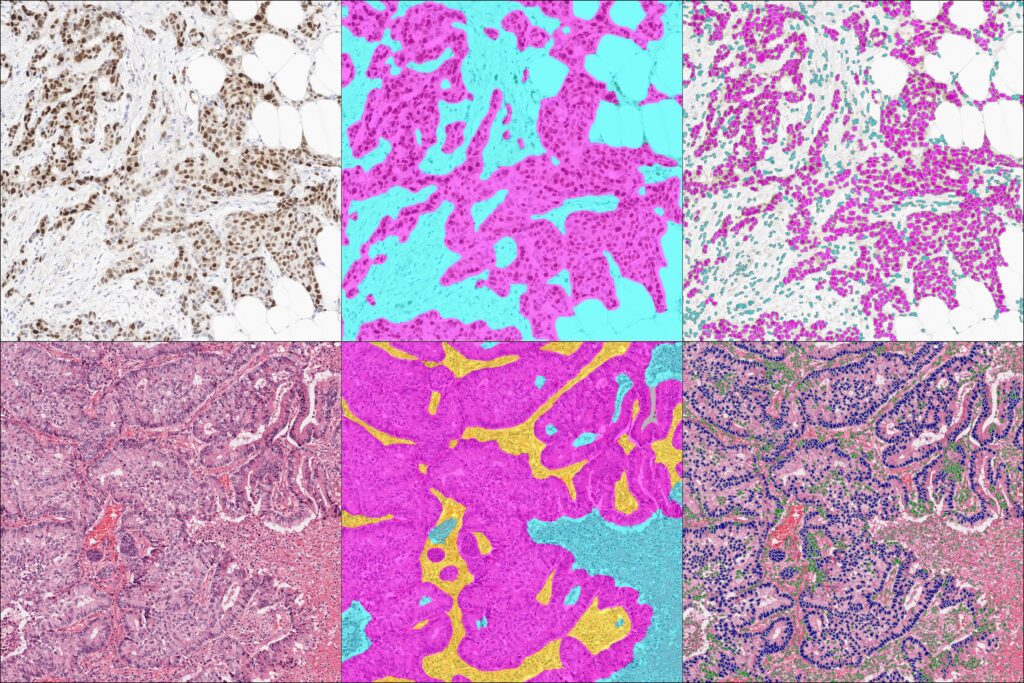
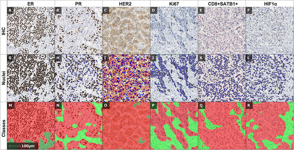
This study by Zilenaite and colleagues evaluated the prognostic value of digital image analysis using HALO on analysis of hormone receptor positive breast cancer IHC biomarkers including ER, PR, HER2, and Ki67 combined with information on tumor heterogeneity and immune response. HALO AI was used for tissue classification to differentiate tumor, stroma, and background (necrosis, artifacts, glass). For quantitative analysis of breast cancer biomarker expression and localization, the Multiplex IHC module of HALO was used. The authors demonstrate that prognostic modeling in hormone receptor positive breast cancer is possible using the computational approach presented here. They also show that the addition of tumor heterogeneity data improved their prognostic model.

Independent Prognostic Value of Intratumoral Heterogeneity and Immune Response Features by Automated Digital Immunohistochemistry Analysis in Early Hormone Receptor-Positive Breast Carcinoma Read More »

Webinar | Digital Pathology in the New Normal: Leveraging HALO® to Investigate a Global Pandemic

21 January 2021 | 8:00 AM – 9:00 AM PST | 11:00 AM – 12:00 PM EST | 4:00 PM – 5:00 PM GMT |
As the world was shutting down in the face of the COVID-19 pandemic, Digital Pathology was thrust to the forefront as a vital option for not only completing existing projects, but more importantly in understanding the disease of COVID-19. Characterizing the mechanisms by which the novel coronavirus attacks the human body, and also understanding the immune system’s response to the disease are critical to guiding treatment and influencing outcomes. Researchers around the world utilized the HALO suite of products to share rare digital pathology slides, collect quantitative pathological data from translational models, characterize gene expression in immune response, and integrate transcriptional data with tissue-based immunoprofiling. This workshop will present some insight on how HALO has been used by researchers at 3 different global institutions to study and characterize COVID-19.

Webinar | Digital Pathology in the New Normal: Leveraging HALO® to Investigate a Global Pandemic Read More »

Spatial Organization of Immune Cells in the Pancreatic Ductal Adenocarcinoma (PDAC) Tumor Microenvironment

28 April 2020 | In lieu of our participation in the now cancelled 2020 AACR Conference, Indica Labs are pleased to bring you webinar presentations from two of our esteemed customers in oncology research and drug discovery, David Krull from GlaxoSmithKline and Dr. David Reiss from Bristol Meyers Squibb, who will present on April 27th and 28th, respectively.   Please join us to hear about how Indica Labs image analysis software and pharma services are being used within their organizations to advance oncology drug development.

Spatial Organization of Immune Cells in the Pancreatic Ductal Adenocarcinoma (PDAC) Tumor Microenvironment Read More »

Digital image analysis improves precision of PD‐L1 scoring in cutaneous melanoma

Viktor H. Koelzer, et al, Nature Medicine, 2018. Immune checkpoint inhibitors have become a successful treatment in metastatic melanoma. The high response rates in a subset of patients suggest that a sensitive companion diagnostic test is required. The predictive value of programmed death ligand 1 (PD‐L1) staining in melanoma has been questioned due to inconsistent correlation with clinical outcome. Whether this is due to predictive irrelevance of PD‐L1 expression or inaccurate assessment techniques remains unclear. The aim of this study was to develop a standardised digital protocol for the assessment of PD‐L1 staining in melanoma and to compare the output data and reproducibility to conventional assessment by expert pathologists.

Digital image analysis improves precision of PD‐L1 scoring in cutaneous melanoma Read More »

A transcriptionally and functionally distinct PD-1+ CD8+ T cell pool with predictive potential in non-small-cell lung cancer treated with PD-1 blockade

Evidence from mouse chronic viral infection models suggests that CD8+T cell subsets characterized by distinct expression levels of the receptor PD-1 diverge in their state

A transcriptionally and functionally distinct PD-1+ CD8+ T cell pool with predictive potential in non-small-cell lung cancer treated with PD-1 blockade Read More »

Scroll to Top