All-In-One Digital Pathology: Compare and Contrast the Tumor Microenvironment of Renal Cell Carcinoma Tissue with Paired, Patient Derived Tumoroids
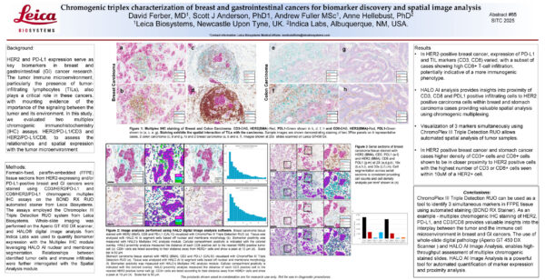
Check out this collaborative poster to learn about a streamlined triplex chromogenic workflow leveraging HALO and HALO AI for analysis of slides assayed with Leica Biosystems’ ChromoPlex III Triple Detection RUO.