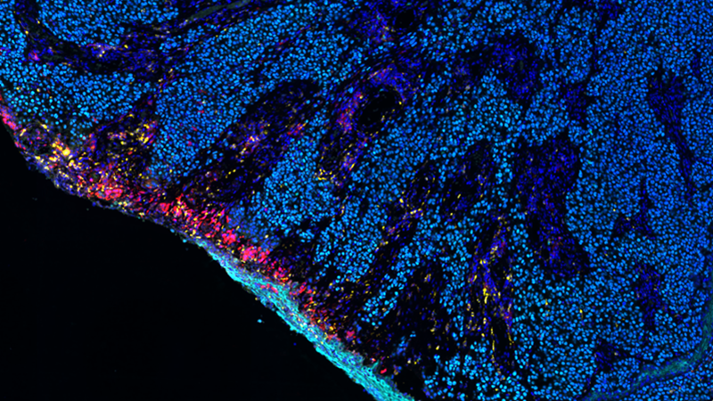
Tumor-specific tumor-resident cytotoxic T cells predict recurrence in stage III melanoma patients treated with adjuvant immunotherapy
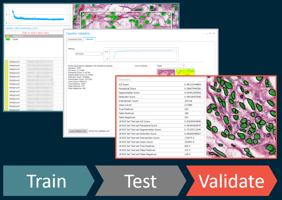
20 January 2022 | In this 60-minute webinar, learn how HALO and HALO AI collaborate to perform tissue and cell segmentation, quantification of T-cell subsets in tumor regions, and spatial analysis .