
Multiplex IHC
Quantify expression of up to five brightfield stains in any cellular compartment – membrane, nucleus or cytoplasm.
Quantify expression of up to five brightfield stains in any cellular compartment – membrane, nucleus or cytoplasm.

25 May 2023 | Following up on our Masterclass Webinar 1 for Multiplex IHC analysis, Masterclass 2 will discuss advanced workflows to help users tackle more challenging brightfield multiplex IHC assays. First, we will demonstrate how advanced cell segmentation and phenotyping options using HALO AI can be combined with the multiplex IHC module to improve image analysis. Two alternative approaches for analyzing multiplex IHC will also be demonstrated including a serial stain workflow to convert brightfield images into pseudo-fluorescence images followed by analysis with Highplex FL and a HALO AI-based approach where cell phenotyping is used to identify cells with co-localization of specific stains. We will discuss advantages, disadvantages and use cases for each workflow.

11 May 2023 | In this 60-minute webinar, we discuss best practices and optimization techniques for advanced quantitative multiplex immunohistochemical (mIHC) image analysis.

In this blog post, you can learn about some of the new features in these modules, where to find the user guides, tutorial videos, and when to expect to upgrade.
HALO®, HALO AI, and HALO Link 3.6 Features and Functionalities Read More »

25 May 2022 | In this 60-min webinar, Learn how HALO and HALO AI are advancing neuropathology research at the UW Medicine Biorepository and Integrated Research (BRaIN) laboratory

10 December 2021 | In this 1-hour webinar, featured speakers will demonstrate end-to-end how to develop a quantitative assay in HALO and transition it into clinical trials using HALO AP.
Bench-to-Bedside Transition of Novel HALO® Assays into Clinical Trials with HALO AP® Read More »

23 September 2021 | DigiBase invites you to attend a Spatial Biology webinar symposium to learn from experts in the field on how digital pathology, high-dimensional multiplexing, spatial and AI analytic tools, and spatial transcriptomic technologies are advancing tissue imaging and analysis.
Spatial Biology: Revolutionizing Tissue Imaging and Analysis Using Spatial profiling Read More »

Myriam Chalabi, et al, Nature Medicine, 2020.
Immunotherapy is known to be effective in late-stage, mismatch repair (MMR) deficient colorectal cancers but not in MMR proficient cancer. This study reports the early results from the NICHE clinical trial that is evaluating treatment of early stage, nonmetastatic preoperative colon cancer with a CTLA-4 inhibitor and a PD-1 inhibitor. Chalabi and colleagues report that the treatment was well tolerated and that a major pathological response was seen in 19/20 patients with MMR deficient tumors and 3/15 MMR proficient tumors. HALO image analysis software was used in this publication for analysis of T-cell biomarkers CD3, CD8, and FOXP3 on chromogenically stained tumor biopsies. First, image registration with HALO was performed of three serial tissue sections. Tissue sections were annotated manually, and quantification of DAB positivity was performed with the Multiplex IHC module and expressed as a function of tumor area. The authors conclude that immunotherapy shows promise to become the standard of care in a defined group of early-stage colon cancers.
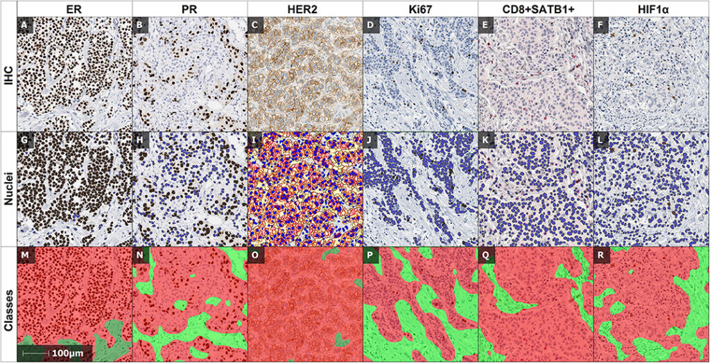
Dovile Zilenaite, et al, Frontiers in Oncology, 2020
This study by Zilenaite and colleagues evaluated the prognostic value of digital image analysis using HALO on analysis of hormone receptor positive breast cancer IHC biomarkers including ER, PR, HER2, and Ki67 combined with information on tumor heterogeneity and immune response. HALO AI was used for tissue classification to differentiate tumor, stroma, and background (necrosis, artifacts, glass). For quantitative analysis of breast cancer biomarker expression and localization, the Multiplex IHC module of HALO was used. The authors demonstrate that prognostic modeling in hormone receptor positive breast cancer is possible using the computational approach presented here. They also show that the addition of tumor heterogeneity data improved their prognostic model.

8 April 2021 | The webinar will cover a workflow for performing stain co-localization analysis in sequential IHC cuts through color deconvolution, slide registration, and cell-type classification, including export using registered coordinates. The resulting co-localization output will be compared and contrasted to true co-expression on a single slide using mIF techniques (Polaris).