Axon Quantification
Quantify axons in nerve cross sections and calculate area, inner and outer diameter, myelin area, and G-ratio.
Axon Quantification Read More »
Quantify axons in nerve cross sections and calculate area, inner and outer diameter, myelin area, and G-ratio.
Axon Quantification Read More »
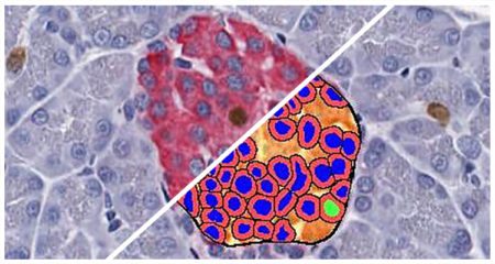
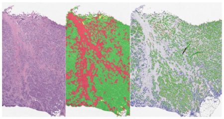
Quantify the area, diameter, perimeter, and number of white spaces per region of interest in brightfield images. Ideally suited for analysis of lipids in brown and white adipose tissue, lipid droplets in liver tissue (steatosis), and alveoli area in lung.
Vacuole Quantification Read More »
The TMA Add-on provides a productivity-enhancing workflow for tissue microarray analysis by enabling automated, high-throughput segmentation and batch analysis of whole slide TMA images.
TMA (Tissue Microarray) Add-On Read More »
Simultaneously analyze an unlimited number of fluorescent dyes and measure object density, area, diameter, and intensity, as well as colocalizations, if applicable.
Object Colocalization FL Read More »
Quantify microglial activation based on length and thickness of microglial processes.
Microglial Activation Read More »
Quantify fiber or membrane positivity for an unlimited number of fluorescent dyes along with fiber diameter, perimeter, and area.
Count and measure pancreatic islets in brightfield images and quantify the islet area and cells positive for up to two islet-specific stains.
Islet Quantification Read More »
Plot cells and objects from one or more images and perform nearest neighbor analysis, proximity analysis, and tumor infiltration analysis.
Analyze serial tissue sections stained with different markers or a single tissue section which has been stained, stripped, and re-stained for multiple markers.
Serial Section Analysis Add-on Read More »
Simultaneously analyze an unlimited number of fluorescent nucleic acid probes on a cell-by-cell basis, measuring spot numbers and area per cell and compartment, and calculated H-scores for each probe.